(214) 420-7611


Joint replacement surgery can be intimidating, especially with concerns about the risks involved. Many people ask, "Is joint replacement a high risk surgery?" This is a crucial question for those dealing with severe joint pain and mobility issues that impact their daily lives. While any surgery has risks, knowing what these risks are and how they can be managed is essential for making informed decisions.
In this blog post, we'll discuss the factors influencing the risk levels of joint replacement surgery. Understanding these aspects will help you have a clearer picture of what to expect, easing any worries and preparing for a successful procedure.
To understand the risks of joint replacement surgery, consider these seven essential factors:
Patient's Overall Health
Your general health is a big factor in determining if joint replacement is a high risk surgery. Conditions diabetes, heart disease, or obesity can increase the risk of complications. For example, someone with uncontrolled diabetes might face higher risks of infection and slower healing. Prior to surgery, your doctor will assess your health and suggest steps to improve your condition, like losing weight or managing diabetes, to reduce risks.
Age
Age can affect how well you recover from surgery. Older adults might have a higher chance of complications due to decreased resilience. However, many older patients successfully undergo joint replacement thanks to advances in medical technology. Older adults need a thorough pre-surgery evaluation to ensure they are fit for the procedure.
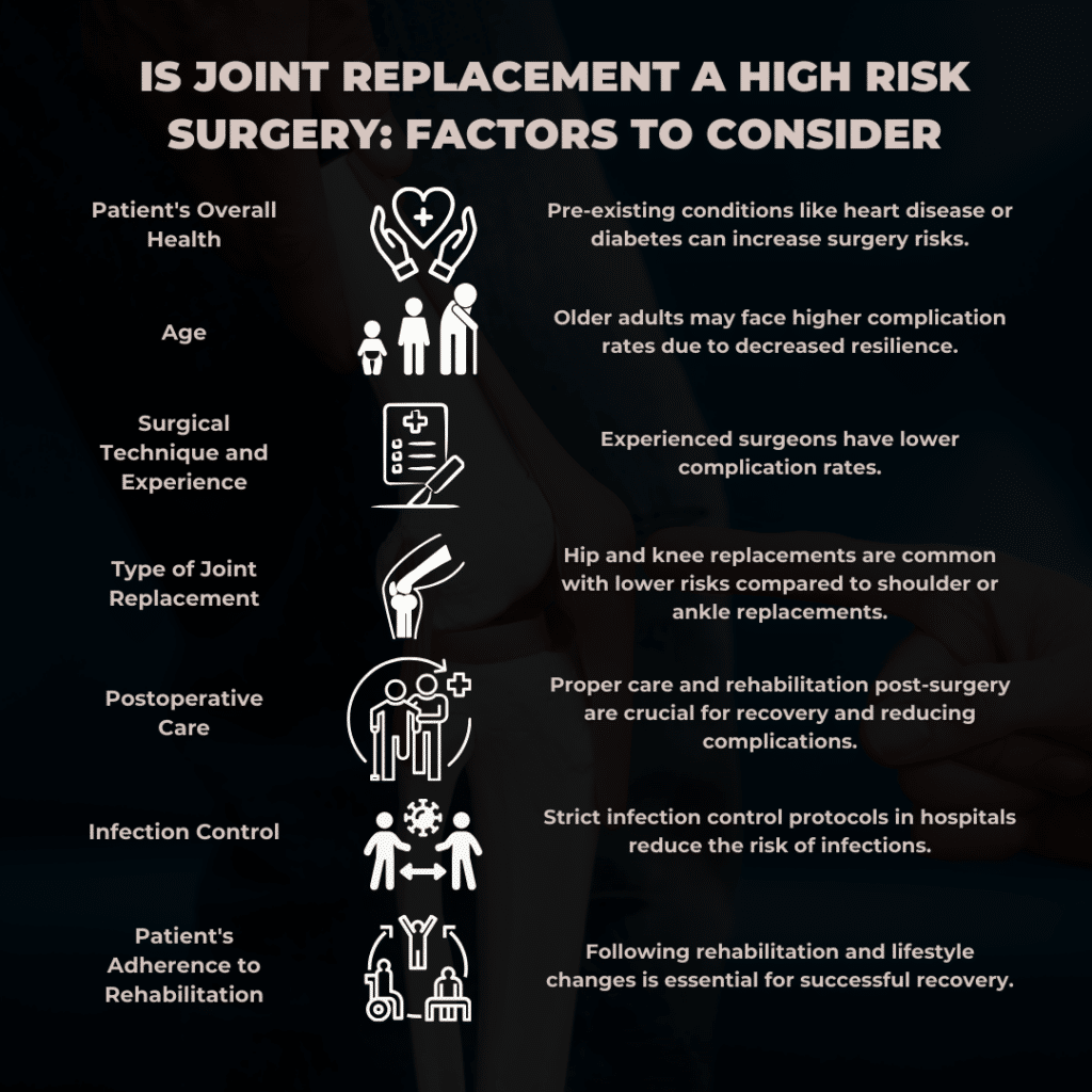
Surgical Technique and Experience
The skill and experience of your surgeon are crucial. Surgeons with a lot of experience in joint replacements tend to have fewer complications. Choosing a well-regarded surgeon can significantly improve the surgery's success. Researching your surgeon's background, asking for referrals, and checking reviews can help you decide whether joint replacement is a high risk surgery for you.
Type of Joint Replacement
The type of joint being replaced also matters. Hip and knee replacements are common and have well-established procedures. On the other hand, shoulder or ankle replacements might have higher risks because they are less common and more complex. Each type of joint replacement comes with its own set of challenges and potential complications.
Postoperative Care
Good care after surgery is vital for a smooth recovery. This includes physical therapy, wound care, and regular check-ups. Proper rehabilitation helps you regain strength and mobility, reducing the chances of complications. It's important to follow your doctor's and physical therapist's instructions closely during recovery.
Infection Control
Infection is a serious risk with any surgery. Hospitals and surgical centers that follow strict infection control protocols, like maintaining sterile environments and monitoring patients properly, greatly reduce this risk. Patients must keep their surgical site clean and following care instructions to prevent infection.
Patient's Adherence to Rehabilitation
Your commitment to following rehabilitation and making lifestyle changes is essential. Sticking to physical therapy and adjusting your daily activities can significantly influence the success of the surgery. Patients who are proactive in their recovery tend to have better outcomes and quicker returns to normal activities.
These factors help answer the question, "is joint replacement a high risk surgery?" By considering each element, you can better prepare and make informed decisions about your health.
Pain is a common concern for anyone considering total knee replacement surgery. Understanding what to expect can help you handle it better. So, how painful is a total knee replacement? Let's break it down.
The pain you experience can depend on how severe your knee problems were before surgery. Those with significant pain and limited mobility may feel greater relief post-surgery, but the initial recovery period can still be challenging.
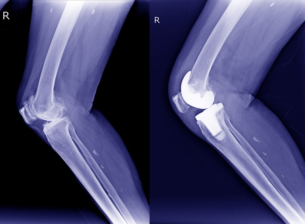
The method your surgeon uses can influence pain levels. Minimally invasive techniques may result in lesser pain and quicker recovery than traditional methods. Discussing different surgical options with your surgeon can help you understand what to expect.
Effective pain management is crucial. Surgeons often use a combination of medications to control pain. These may include local anesthetics, oral painkillers, and sometimes nerve blocks to numb the area. Post-surgery, you will receive a pain management plan tailored to your needs.
Physical therapy is essential for recovery. It helps regain strength and mobility, but the exercises can be painful initially. Following your therapist's instructions can help manage this pain effectively. The more committed you are to your rehabilitation, the better your recovery will be.
Everyone's pain tolerance is different. What might be very painful for one person could be more manageable for another. Communicate with your medical team about your pain levels so they can adjust your treatment accordingly. It's important to stay ahead of the pain rather than letting it become severe.
Swelling is a typical part of recovery and can contribute to discomfort. Ice packs, elevation, and prescribed medications can help reduce swelling and pain. Keeping the swelling under control is key to a more comfortable recovery.
Although rare, complications like infections or blood clots can cause additional pain. Following your doctor's post-surgery care instructions can minimize these risks. Awareness of the signs of complications and addressing them early can make a big difference in your recovery.
In summary, while a total knee replacement can be painful, effective pain management strategies and proper postoperative care can help manage and reduce this pain. Understanding these aspects will help you prepare better for the recovery journey and address concerns about "is joint replacement a high risk surgery."
Is total knee replacement a high risk surgery? Generally, the risks are similar to those of other joint replacements, but specific factors can make a difference.
Both joint replacement and total knee replacement surgeries share common risks such as infection, blood clots, and complications from anesthesia. However, knee replacements are often more straightforward due to their prevalence and the extensive experience surgeons have with these procedures.
Therefore, while both surgeries carry risks, knee replacements' specific nature and commonality often result in slightly lower risk levels. For many, the question "is joint replacement a high risk surgery" may have a slightly different answer when considering knee replacements.
Recovery after knee replacement surgery involves several stages. Here's what you can generally expect:
Most patients stay confined in the hospital for 2-3 days. You'll start with gentle movements and physical therapy exercises during this time. The medical team will monitor your progress and recovery and manage your pain, which is a crucial part of addressing "is joint replacement a high risk surgery."
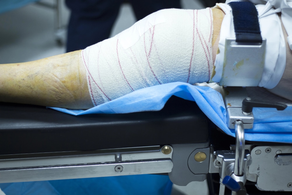
Pain will be managed with medications. It's crucial to follow your doctor's instructions to stay ahead of the pain. Effective pain management helps you start moving and participating in therapy sooner.
You'll likely begin walking with a walker or crutches within a day after surgery, preventing blood clots and improving circulation. Early movement is important for a successful recovery.
Continue physical therapy to regain strength and mobility. Exercises will gradually become more intensive. Your therapist will guide you through a personalized plan to ensure steady progress.
Keep the surgical site clean and dry. Follow your doctor's instructions for wound care to prevent infection. Proper wound care is paramount for healing and preventing complications.
You'll be encouraged to move around and perform light activities to aid recovery. Avoid strenuous activities until your doctor says so. Gradually increasing your activity level helps build strength and flexibility.
Full recovery can take several months. Consistent physical therapy is vital for regaining full function. Staying committed to your rehabilitation plan is key to achieving the best outcome.
To support your new joint, incorporate exercises that strengthen your knee and maintain a healthy weight. Long-term success hinges on making healthy lifestyle choices and staying physically active.

Regular check-ups with your surgeon make sure your knee is healing properly and allows for adjustments in your recovery plan. Keeping up with follow-up visits is important for monitoring your progress and addressing any issues.
Understanding these stages will help you set realistic expectations and prepare for the journey ahead.This comprehensive approach addresses the question, "is joint replacement a high risk surgery," by outlining the steps to a successful recovery.
Joint replacement surgery, while not without risks, offers significant benefits for those suffering from severe joint pain and mobility issues. Patients can confidently navigate this process by understanding the key factors and preparing adequately.
If you're considering this procedure, discussing your concerns with a medical professional is paramount so you make the best health decision. Sunnyvale Sports Medicine and Orthopedic Center can provide expert advice and care throughout your joint replacement journey.
How do you make a shoulder injury heal faster? The short answer is to find out what is causing the pain, protect the joint, and follow the right rehab plan. A mild sprain may feel better within weeks. A tendon tear or frozen shoulder can take much longer.

Your shoulder can move in almost every direction. That wide range of motion is useful, but it also makes the joint easier to injure.
The upper arm bone sits in a shallow socket in the shoulder blade. Muscles, tendons, ligaments, and the labrum help hold everything in place.
The rotator cuff is especially important. These muscles and tendons help you lift and turn your arm.
Understanding these parts can also make it easier to explain a shoulder injury to your healthcare provider.
Shoulder pain can start after a fall, heavy lifting, sports, or repeated overhead use. Wear over time can also cause problems.
Healing time depends on what is injured and how severe the damage is.
Shoulder impingement and bursitis often hurt when you reach overhead or lift your arm.
Many cases improve within a few weeks to a few months. Rest from painful activities and guided exercises often help.
Surgery is not usually the first step. It may be considered when pain continues after a full course of non-surgical care.
A rotator cuff tear can be partial or complete. You may notice weakness, pain at night, or trouble lifting your arm.
The American Academy of Orthopedic Surgeons says many patients improve without surgery.
Surgery may be discussed when there is:
The labrum is a rim of tissue around the shoulder socket. It helps keep the joint stable.
A SLAP tear is one type of labral tear, not another name for every labral injury.
Smaller tears may improve with rest and physical therapy. Surgery may be needed when pain or instability continues after rehab.
Adhesive capsulitis is better known as frozen shoulder. It causes pain and a gradual loss of movement.
Recovery is often slow. According to AAOS:
Most people start with non-surgical treatment. Surgery is rarely needed.
The AC joint sits where the collarbone meets the top of the shoulder. A fall directly onto the shoulder can injure it.
Johns Hopkins Medicine reports that a Grade 1 sprain may recover in about 10 to 14 days. A Grade 3 injury may take around 6 to 8 weeks to regain motion and function.
Most Grade 1 to 3 injuries can be treated without surgery. Severe or lasting problems may need an orthopedic review.

A shoulder fracture can involve the collarbone, upper arm bone, or shoulder blade.
Common symptoms include pain, bruising, swelling, and trouble moving the arm.
Many fractures heal without surgery. Bone healing often takes about 6 to 12 weeks, though strength and movement can take longer to return.
Surgery may be needed when the bone is badly displaced or unstable.
Shoulder arthritis is a long-term joint problem rather than an acute injury.
Osteoarthritis, rheumatoid arthritis, and post-traumatic arthritis have different causes. Treatment may include exercise, medication, injections, and activity changes.
Joint replacement may be considered when pain is severe, and other treatments no longer help.
| Condition | Typical Recovery | When Surgery May Be Considered |
| Impingement or bursitis | Weeks to months | Pain continues after non-surgical care |
| Rotator cuff tear | Several months | Large tear, trauma, weakness, or lasting pain |
| Labral tear | Several weeks or longer | Ongoing pain or repeated instability |
| Frozen shoulder | Months to years | Severe stiffness that does not improve |
| AC joint sprain | About 10 days to 8 weeks | Severe injury or lasting symptoms |
| Shoulder fracture | Often 6 to 12 weeks | Unstable or badly displaced fracture |
There is no safe way to force an injured shoulder to heal overnight. The goal is to give the tissue what it needs while keeping the joint from getting too stiff.
A few simple steps can help:
Physical therapy is often part of shoulder treatment.
A therapist may work on your range of motion first. Strength exercises can be added as the shoulder becomes less painful.
Treatment often focuses on the rotator cuff and shoulder blade muscles. The exact plan should match your diagnosis.
Sharp or increasing pain is a sign to stop and speak with your provider.

Not every patient needs the same treatment. Your plan may include:
RICE stands for rest, ice, compression, and elevation. It can be useful for short-term care after some injuries.
Still, keeping the shoulder completely still for too long can lead to stiffness. Safe movement is usually added as symptoms settle.

Physical therapy should start at the right time, not simply as soon as pain begins.
See a medical provider first after a hard fall, dislocation, or other major injury. You should also get prompt care if the shoulder looks deformed or you cannot lift your arm.
For many overuse injuries, therapy can begin once it is safe to move the joint. Early guided movement may help restore function and reduce stiffness.
At Sunnyvale Sports Medicine and Orthopedic Center, treatment begins by finding the source of your shoulder pain. Book an appointment if pain is affecting your sleep, work, exercise, or daily routine.
A mild Grade 1 AC joint sprain may improve within 10 to 14 days. A Grade 3 injury may take around 6 to 8 weeks. Return to sports can take longer, depending on pain and strength.
Common exercises include shoulder blade squeezes, gentle arm raises, and external rotation work. A therapist may also use cross-body stretches. The best exercises depend on what is causing your pain.
Pain and function may improve over several weeks to several months with therapy. A full-thickness tear may not reattach to the bone on its own. Non-surgical care can still reduce pain and improve how the shoulder works.
Surgery may be considered when pain or instability continues after physical therapy. Repeated dislocations can also be a reason to repair the labrum. The tear type, activity level, and symptoms all matter.
RICE means rest, ice, compression, and elevation. It may help control pain and swelling during the early stage of some injuries. It should not replace medical care when symptoms are severe.
Shoulder injuries do not all heal on the same schedule. The right treatment depends on the tissue involved and the extent of the damage. If shoulder pain is not improving, book an appointment with Sunnyvale Sports Medicine and Orthopedic Center to find the cause and plan your next step.
Joint pain can be debilitating. It can affect your quality of life and limit your activities. If you're considering joint replacement, you might wonder, "Is joint replacement a major surgery?" Understanding this procedure is crucial for making an informed decision about your health.
Let's explore the essential aspects of joint replacement surgery in this blog. We'll discuss whether joint replacement is considered major surgery, the success rates, potential risks, and other critical factors.
By the end of this post, you'll understand what to expect and be better prepared to decide if this procedure is right for you.
Keep reading to discover key facts and insights about joint replacement surgery.
Anyone considering joint replacement must understand whether it is a major surgery. Here are five key facts to help clarify the nature of joint replacement surgery.
1. Involves Significant Surgical Procedures
Joint replacement involves the removal of damaged or diseased parts of a joint, which are then replaced with artificial components. This complex process requires precise surgical techniques, often making it a major surgery. The intricacies of removing and replacing joint parts highlight why joint replacement is considered a major surgery.
2. Requires General or Regional Anesthesia
Patients undergoing joint replacement typically need general or regional anesthesia. This ensures they are comfortable and pain-free during the procedure. Anesthesia carries risks and adds to the surgery's complexity, underscoring that joint replacement is a major surgery.

3. Extensive Recovery Period
Recovery from joint replacement surgery can take some time. Patients often need several weeks to months to recover fully. Physical therapy is typically required to regain your strength and mobility, which adds to the overall impact of the surgery. This extended recovery period is another reason why joint replacement is considered a major surgery.
4. Potential Complications
Like any major surgery, joint replacement carries risks of complications. These can include infection, blood clots, and issues with the artificial joint itself. These potential complications further emphasize that joint replacement is a major surgery. Recognizing the risks involved is crucial for anyone asking, "Is joint replacement a major surgery?"
5. Long-Term Benefits
Despite being a major surgery, joint replacement can offer significant long-term benefits. Numerous patients notice a decrease in pain and an improvement in mobility, significantly enhancing their quality of life. This positive outcome often justifies the surgery's extensive nature, proving that the benefits can be substantial while it is a major surgery.
So, is joint replacement a major surgery? With all these factors considered, it certainly fits the criteria. These key facts can help you decide about joint replacement surgery. It's essential to weigh the complexities and benefits to determine if this procedure suits you.
The success of joint replacement surgery is a crucial factor for those considering this procedure. Knowing the outcomes can help patients feel more confident in their decisions. Here are five key facts about the success rates of joint replacement surgery, mainly focusing on total knee replacement (TKR).
High Success Rates for All Ages
Studies show total knee replacement (TKR) has a high success rate, even for individuals aged 65 and older. Pain relief and improved mobility and function have been reported from six months to ten years post-surgery. This indicates that joint replacement is a major surgery with long-term benefits.
Patient-Reported Outcomes

For older adults, TKR results in significant improvements in pain, function, and quality of life. These outcomes are critical as they directly impact daily activities and overall well-being. Satisfaction levels among patients aged 65 and over are high, further supporting the procedure's success. The question often arises, "Is joint replacement a major surgery?" Given these outcomes, it’s clear that the procedure has a significant impact.
Factors Affecting Success
The procedure's success depends on several factors. Patient health, surgeon experience, and post-surgery care all play vital roles. Ensuring proper rehabilitation and following medical advice can enhance recovery and long-term results. This further underscores why joint replacement is a major surgery. Comprehensive preoperative planning and postoperative care are crucial to achieving the best possible outcomes.
Improved Quality of Life
Health-related quality of life significantly improves after TKR. Studies report notable enhancements in both physical function and overall well-being. These improvements are seen consistently across various age groups, making joint replacement a reliable solution for managing severe joint issues. Is joint replacement a major surgery? The improvements in quality of life affirm that it indeed is.
Long-Term Satisfaction
Satisfaction with TKR remains high, with many patients reporting positive outcomes even years after the procedure. This satisfaction strongly indicates the surgery’s effectiveness in alleviating pain and restoring mobility. Understanding these success rates helps answer the question, "Is joint replacement a major surgery?"
These key points highlight joint replacement surgery's effectiveness and high success rates. For anyone considering this procedure, these insights provide valuable information to help make an informed decision.
While highly beneficial, joint replacement surgery comes with its own risks. Here, we discuss whether joint replacement is a high-risk surgery and what potential complications may arise.
Surgical Risks
Joint replacement is a major surgery; like any such procedure, it carries risks. These include infection, blood clots, and adverse reactions to anesthesia. While these risks are relatively low, they are essential to consider.
Infection

As with any surgery, infection is a potential risk with joint replacement. The surgical site can become infected, which may require additional treatment or even another surgery. To minimize this risk, surgeons follow strict sterilization protocols, and patients may receive antibiotics before and after surgery.
Blood Clots
After joint replacement surgery, blood clots can form in the legs (deep vein thrombosis) or lungs (pulmonary embolism). To minimize this risk, patients are prescribed blood thinners and strongly encouraged to start moving as soon as possible following surgery.
Implant Issues
Over time, the artificial joint can wear out or become loose. This may necessitate a revision surgery to replace the worn-out components. While modern implants are designed to last longer, this risk is still present and should be discussed with your surgeon.
Long-Term Risks
Other long-term risks include nerve damage, joint stiffness, and reduced range of motion. Although these complications are rare, they can impact the overall success of the surgery. Patients must be aware of these potential issues and discuss them with their healthcare provider.
Is joint replacement a major surgery? Yes, and while it is a major surgery with inherent risks, advancements in medical technology and surgical techniques have significantly reduced these risks. Most patients experience successful outcomes with proper preoperative and postoperative care.
If you are considering joint replacement, understanding these risks can help you make a sound decision. So, is joint replacement a major surgery? Considering all the factors, it certainly fits the criteria.
Joint replacement surgery can significantly enhance the quality of life for people who suffer from severe joint pain. Understanding the complexities and benefits of the surgery is crucial for making an informed decision.
While joint replacement is a major surgery, it offers substantial long-term benefits, including pain relief and improved mobility. You can determine if this procedure is right for you by weighing the risks and benefits.
Consulting with a trusted medical professional is essential if you're considering joint replacement. Sunnyvale Sports Medicine and Orthopedic Center provides comprehensive diagnostic and treatment services to help manage joint issues and improve your overall well-being.
Shoulder instability is a condition that can significantly impact your daily life. It occurs when the structures surrounding the shoulder joint do not work as they should, leading to frequent dislocations and a persistent feeling of the shoulder being loose.
Many people may underestimate the importance of addressing this issue and understanding what happens if shoulder instability is left untreated, thinking it will be resolved independently. However, the consequences of neglecting shoulder instability can be severe.
If shoulder instability is not addressed, the condition can eventually worsen, leading to more frequent dislocations, increased pain, and potential long-term damage to the shoulder joint.
In this article, let's explore the seven significant consequences of untreated shoulder instability, providing valuable insights into why early intervention is crucial.
Understanding what happens if shoulder instability is left untreated can help you make informed decisions about your health. Continue reading to learn about the significant impacts and how they can affect your overall quality of life.
When shoulder instability is left untreated, several serious consequences can arise. Understanding these outcomes emphasizes the importance of seeking timely medical intervention.
The shoulder joint remains unstable without proper treatment, leading to frequent dislocations. Each dislocation causes further damage to the ligaments, tendons, and muscles, exacerbating the instability.
Persistent instability often results in chronic pain. This pain can limit daily activities and reduce the overall quality of life. This makes it undeniably challenging to perform even simple tasks like lifting objects or reaching overhead.

Repeated dislocations can lead to significant damage to the shoulder joint. Over time, this damage can cause arthritis, characterized by joint pain, swelling, and reduced mobility.
The muscles surrounding the shoulder can weaken due to repeated dislocations and lack of stability. This weakness further contributes to instability, creating a complex cycle that cannot be broken without intervention.
Untreated shoulder instability often results in a decreased range of motion. Activities that require shoulder movement become increasingly tricky, limiting participation in sports, work, and daily activities.
Frequent dislocations can damage the nerves around the shoulder joint. Nerve damage can cause numbness, tingling, and loss of sensation in the shoulder and arm, further complicating the condition.
Chronic pain, limited mobility, and ongoing medical issues can significantly impact mental health. Anxiety, depression, and a sense of helplessness are common among those dealing with untreated shoulder instability.
Addressing shoulder instability early is crucial to prevent these severe consequences. With timely intervention, knowing what happens if shoulder stability is left untreated can restore stability, reduce pain, and improve shoulder joint function.
An unstable shoulder can manifest in several ways, often causing discomfort and difficulty with everyday activities. Here are the common sensations and symptoms associated with an unstable shoulder:
An unstable shoulder may frequently dislocate or partially dislocate (subluxation). This sensation can feel like the shoulder is slipping out of place and can happen during simple movements or activities. These episodes can be sudden and unpredictable, making it difficult to trust the stability of your shoulder during any activity.
Pain is a common symptom of shoulder instability. It can cause sharp pain during a dislocation or constant dullness due to ongoing instability and stress on the shoulder joint. Pain may also increase with certain movements, such as lifting or reaching, and can sometimes extend down the arm.

Many people with shoulder instability experience weakness in the shoulder and arm. This weakness can make lifting objects challenging or performing tasks requiring strength. The lack of stability can cause muscles to compensate inappropriately, leading to fatigue and further weakness.
A feeling of looseness or that the shoulder might "give way" during activities is typical. This sensation can create anxiety about using the shoulder for certain movements. This can be particularly concerning during activities that involve overhead motions or bearing weight on the shoulder.
Some individuals may notice clicking or popping sounds when moving their shoulders. These sounds can be a sign of instability and underlying joint issues. The noises may be accompanied by a sensation of catching or locking, adding to the discomfort and unpredictability of shoulder movement.
Shoulder instability can result in a limited range of motion and difficulty performing tasks requiring full shoulder mobility. This restriction can impact daily activities, including dressing, reaching for items on high shelves, and participating in sports and exercises.
Repeated dislocations or subluxations can cause swelling and inflammation in the shoulder joint. This can lead to stiffness and increased pain, further limiting the shoulder's functionality and comfort.
Understanding these symptoms can help identify shoulder instability early. Recognizing what happens if shoulder instability is left untreated is vital for seeking timely medical care and preventing further complications. Shoulder instability can be managed better when diagnosed early and treated promptly, improving the quality of life for those affected.
Shoulder instability can be temporary or permanent, depending on several factors, such as the cause, severity, and treatment.
In some cases, shoulder instability is temporary and can be resolved with proper treatment. Physical therapy and rehabilitation exercises can strengthen the muscles around the shoulder, improving stability and function.
In other instances, shoulder instability may become a long-term issue. This can occur if the condition is severe, significant joint damage has occurred, or it is not treated promptly. Surgical intervention may be recommended to restore stability in such cases.
The likelihood of shoulder instability being permanent depends on factors like age, activity level, and the number of previous dislocations. Younger individuals or those involved in high-risk sports may have a higher chance of recurring instability.
Understanding the potential permanence of shoulder instability highlights the importance of early intervention. Recognizing what happens if shoulder instability is left untreated can motivate individuals to seek the necessary care to avoid long-term issues.
Shoulder instability surgery aims to restore stability to the shoulder joint, and its success rate is generally high.
Over 80% of patients report good or excellent outcomes after shoulder instability surgery. This indicates a significant improvement in stability, function, and pain reduction. Clinical scores used to measure outcomes often show marked improvement post-surgery, reflecting the procedure's effectiveness.

Despite the high success rate, the recurrence rate is still 5.1% to over 20%. Factors such as the patient's age, activity level, and the number of previous dislocations can influence this. Younger patients and those engaged in high-impact sports may have a higher likelihood of recurrence, necessitating careful post-surgical management and possible lifestyle adjustments.
Patient satisfaction with shoulder instability surgery is typically high. Over 80% of patients return to their previous activity levels, indicating that the surgery effectively restores shoulder function. High patient satisfaction rates are often attributed to significant pain reductions and improvements in the shoulder's range of motion and strength.
The long-term benefits of shoulder instability surgery include sustained stability and improved shoulder joint function. While some patients may experience degenerative changes over time, these do not necessarily impact the overall functional outcomes. Regular follow-up and physical therapy can help maintain these benefits and prevent further complications.
The success of shoulder instability surgery also depends on the rehabilitation process. A well-structured physical therapy program is crucial for strengthening the shoulder muscles and improving joint stability. Adherence to post-operative care instructions significantly enhances recovery outcomes and minimizes re-injury risk.
Understanding these success rates and associated factors helps set realistic expectations for those considering surgery. Knowing what happens if shoulder instability is left untreated can encourage patients to explore surgical options if non-surgical treatments are insufficient.
Shoulder instability is a serious condition that can lead to numerous complications if left untreated. Chronic pain, frequent dislocations, joint damage, and even mental health issues are just among the many potential consequences. Recognizing what happens if shoulder instability is left untreated can motivate individuals to seek early medical intervention, improving their chances of successful treatment and recovery.
Shoulder instability can be challenging, but at Sunnyvale Sports Medicine and Orthopedic Surgery Center, our dedicated team is experienced in knowing what happens if shoulder instability is left untreated and providing comprehensive care to restore stability and alleviate pain. If you're showing signs of shoulder instability, don't wait to seek expert advice and explore your treatment options.
Labral tears in the hip might cause painful locking or clicking of the hip, reduced range of motion, and disruption to daily activities and sports. The acetabular labrum aids in the stabilization of the hip joint, especially during hip motion. The fibrocartilaginous structure extends the acetabular rim and offers a suction seal around the femoroacetabular interface.
Hip labral tears do not heal easily so that surgery may be necessary in many cases. Surgery aims to improve hip stability, function, and range of motion, decrease hip pain, and prevent additional damage to the hip joint. But is labrum repair a major surgery?
A hip labral tear is a type of injury that impacts the hip's labrum. This tough, crescent-shaped cartilage structures the rim and lines of the hip socket (called the acetabulum), which is found in the pelvic bone. Also called the acetabular labrum, it shouldn't be confused with the shoulder labrum, which is a similar structure known as the glenoid labrum.
The hip functions like a ball-and-socket joint, with the head of the femur (the thighbone) forming a "ball" that fits into a "socket" known as the acetabulum found in the pelvis. Both the ball and socket have smooth articular cartilage covering them. The labrum is an extra, specialized piece of cartilage that runs along the socket's rim to provide a suction seal and stability to your hip joint. It helps absorb shock and spread pressure during hip movement, ensuring smooth motion. The hip labrum may become torn or detached from the acetabular socket for various reasons.

A torn labrum can be caused by:
·Repetitive motions over time lead to "wear and tear."
·A traumatic injury is very common in athletes who carry out repeated hip flexions and sudden impact on their hips, such as hockey players, runners, soccer players, and football players.
·Individuals with other conditions of the hip can be especially susceptible to labral tears. These include:
·Hip impingement (also called femoroacetabular impingement) is a condition in which abnormal contact between the hip's ball and socket is due to a deformity of the acetabulum or the femoral head.
·Osteoarthritis of the hip, a cartilage degeneration, can lead to rough, bone-on-bone contact in the hip.
Hip labral tear symptoms can include:
·Deep pain in the groin or the buttocks on the side of the injured hip's side.
·A sound or feeling of locking or clicking when your hip is in motion.
·Hip pain, particularly while it rotates in specific directions.
·Stiffness while moving your hip.

Labral tears are hard to diagnose, partially due to the many muscles and other structures near the hip joint. They are usually misdiagnosed as common groin strains, and it isn't uncommon for the diagnosis to be missed for several months after your labrum is torn.
A sports medicine physician or orthopedist will perform a physical exam and consider your symptoms. If your doctor thinks you might have a torn labrum, they will order hip imaging. This may include X-rays, but a hip MRI is the best way to confirm the diagnosis.
Your doctor might advise hip arthroscopy if you have a severe tear or if a nonsurgical procedure won't provide adequate relief. This type of arthroscopic procedure is usually done on an outpatient basis. The procedure is small incisions about half a centimeter around the hip using specialized instruments to either debride (clean) remove the injured labrum or reattach it to the socket. Following the surgery, you'll be discharged on crutches and use them for 2-6 weeks. This length of time is identified on a case-by-case basis. Post-surgical physical therapy will improve your muscle strength around the hip and range of motion.
Whether treated nonsurgically or surgically, recovery from a torn hip labrum can last up to six weeks. Based on the extent of the injury, competitive athletes may return to their sport between two and six months after the injury.

During surgery, a doctor might perform:
Labral tear repair. Your doctor will reattach the torn labrum to the socket (acetabulum) with small plastic "anchors" and sterile thread.
Debridement. This technique involves trimming or smoothing the frayed area of the labrum. The goal is to remove damaged, loose, and pain-generating tissue.
Other repairs. Hip problems are usually intertwined, so it's typical for a surgeon to repair your hip labrum and deal with other problems during the same surgery. For instance, the surgeon might repair the labrum and shave down bony abnormalities that lead to hip impingement.
Other repairs may be made during surgery. Pre-operative medical imaging cannot always reveal everything wrong in a joint. A surgeon might confirm or discover other hip issues for the first time during surgery.
Labral reconstruction utilizes a graft—tissue from another part of the body or a donor—to completely replace the torn labrum. This procedure occurs when the labrum is so damaged that it can't repaired, but the hip joint is otherwise healthy.
The type of surgery advised will be based on the severity of your injury or condition and other underlying conditions that must be addressed.

People considering hip labrum repair should consider how they cope with post-surgical recovery.
Recovery often requires:
·Taking pain medication for 1-3 days
·Using crutches (in some instances, a walker might be needed initially)
·Dedicating time to physical therapy
Many patients are eager to learn about their anticipated recovery from labral tear hip surgery. That said, it's crucial to remember that each treatment and recovery will differ for everyone. Postoperative recovery times differ depending on the severity of your hip labrum tear, related chronic conditions, and other factors. Nevertheless, most patients should expect to utilize crutches for the first two weeks after their hip labrum surgery. Some patients may need six months to recover from hip labrum surgery fully. No matter how long it takes, your doctor will track your progress during hip labral tear surgery recovery and suggest a rehab strategy, including torn labrum hip exercises when appropriate.
People with sedentary jobs often return to work 1-2 weeks after arthroscopic hip labrum surgery. Prolonged sitting isn't recommended, so these individuals will be encouraged to stretch and move intermittently across the day.
People with labor-intensive jobs, like those requiring heavy lifting or standing for long periods, may need to wait up to 6 months following arthroscopic hip surgery before returning to work.
Generally, an individual who has undergone arthroscopic hip surgery can return to moderate activity, like jogging, after three months, although some people may be required to wait longer. For high-hip-intensity sports, such as ballet, football, rowing, and lacrosse, a 5—to 6-month timeframe for recovery is typical following hip arthroscopy. This time frame depends on many factors, such as the type of surgery performed, the person's fitness level, and the sport itself.
Arthroscopic surgery is virtually always an outpatient procedure, and you might expect to go home within hours of surgery. Most people can walk with crutches almost immediately after the procedure. Our pain management experts ensure you have the medication to stay comfortable during recovery.
Doctors suggest using crutches for 2-3 weeks, and your surgeon books a follow-up appointment for two weeks after surgery to take off the stitches and confirm that your incision wounds are healing normally. Your surgeon also checks your hip and asks you to rotate your leg gently in various directions to assess its range of motion following surgery.
Our doctors suggest starting physical therapy in the days following surgery. Physical therapy helps rebuild hip muscle flexibility, range of motion, and strength.
At Sunnyvale Sports Medicine, we specialize in the latest conservative care treatment options and arthroscopic surgical techniques to treat hip labral tears. If you or a loved one are being held back by the pain and discomfort of a hip labral tear, come for a consultation to learn more about the latest treatment alternatives. Our team is committed to allowing patients to achieve their active lifestyle goals without limitations.
You've recovered from knee replacement surgery and are ready for a relaxing vacation. However, certain types of joint implants can trigger airport security alarms. Do knee replacements set off metal detectors? Well, it is best to expect security screening to discover implants.
Airport security screening measures' sensitivity has immensely increased during the past decade, but several reports have examined how this affects patients with artificial joints. Over 90 percent of implanted total hip and knee arthroplasty devices will trigger airport metal detectors.
With varying levels of security in airports worldwide and varying clearance provided by TSA Precheck or Clear, patients with a new joint may set off the detector in one city but not another. Joint replacement surgery is increasingly in demand as national healthcare systems confront an ever-aging population. This, in turn, affects air travel; more specifically, unwanted and significant inconveniences are caused to travelers going through airport security.
Here's what you need to know to make your security trip smooth.

Of the 143 patients suffering from hip replacements who traveled by air, 120 (84 percent) reported setting off the alarm and required wanding with a handheld detector.
Twenty-five of these patients stated subsequently having to undergo further inspection, such as additional wanding, being patted down, and, in two instances, having to undress in a private room to present the incision.
Ninety-nine (69 percent) of the 143 patients reported that the prosthetic joint caused an inconvenience while traveling.
In another similar study that was designed to characterize the airport metal detection's efficacy of total knee prostheses, the delays encountered, any inconvenience this might have caused, and the role of implant identification cards, 97 total knee arthroplasty recipients stated going through an airport metal detector, with 70 setting off the alarm a mean of 3 times (range, 1 to 36). A single-knee prosthesis activated airport security alarms more than 83 percent of the time and increased patient inconvenience. Patients must be informed about this chance and be prepared to present documentation of their prostheses.
According to the American Society for Hip and Knee Surgeons, a card or note from your physician is optional for the identification of these types of implants. However, based on a study examining the effects of the increase in air travel for aging patients, looking specifically at the impact of artificial joints on travel, both airport security officers and patients concluded that joint replacement implant identification cards streamline airport security checks and decrease the need for more invasive searches at airport security.
However, even with such a note, a traveler may still experience some level of inconvenience and should be cautioned beforehand to prepare for travel delays and give themselves extra time at the airport. If a person has a metal implant, it is advisable to inform the Transportation Security Administration (TSA) officer before screening begins that you have had a hip or knee replacement and to point to the location of the implant.

Passengers can utilize the TSA's Notification Card to communicate discreetly with security officers. Showing this card or telling the TSA officer won't exempt a passenger from the screening. Instead, the passenger will be instructed to undergo the body scan machine. Many individuals prefer to be screened by the body scan to lessen the likelihood of a pat-down being necessary.
Because of the necessity for airport security, passengers with artificial joints will likely experience more inconvenience when traveling. To ease discomfort, it is recommended that security personnel be informed in advance.
Although only some people are asked for proof of their surgery, the orthopedic surgeon can give a card that confirms you're using an artificial joint. These cards identify the holder as having a joint replacement and state that the implant may set off metal detection devices. Keep your joint replacement card when you travel in case you need to present proof of your implant.
Though the metal implant can cause inconvenience by triggering the alarm, it does not cause any additional harm to your body. Metal detectors use an electromagnetic field that contains no harmful radiation. However, if you do trigger the metal alarm, X-ray machines are increasingly used in many airports, and they use very low levels of ionizing radiation.
It's perfectly safe to kneel on a knee replacement once the incision is completely healed. However, most people find it relatively uncomfortable. If you're going to kneel, it's recommended that you put a pad or pillow beneath your knee to make it more comfortable.

Currently, there are no regulations in security screening for those who have implants, including plates, screws, nails, and joint replacement in their body.
The advice is that there is a good chance that they will set off the metal arch. If that happens, you will have to undergo a secondary screening (X-ray machine; handheld detector) irrespective of whether you have an ID card, a clinic note, or even a scar in that region.
The sensitivity of the metal detectors ranges from 1 to 15, but in some countries, it is 0 to 99. It is normally kept at around 5; however, the sensitivity will increase with an increased security threat. Suppose the sensitivity of the metal detectors is increased.
In that case, they will detect smaller amounts of metal in your body, which could be why some arch metal detectors detect your implant. The handheld metal detectors are usually more sensitive than the arch ones, and they find the implants more easily.

Depending on the type of implant used, implants with a higher iron content are easily detected. However, the detection rate for modern implants is quite variable. The cobalt-chromium alloy commonly used in joint replacement is easily detected compared to titanium and stainless-steel implants.
The heavier the metal implant used in the body, the easier it is to detect. Depending on the type of joint replacement used, the weight could range from 130 g to 3 kg. There is a good chance of setting off the alarms if you have a joint replacement, have a rod in the bone, or have a big plate with more than ten holes.
There is conflict in reports based on your body mass index, with some data showing that if you weigh a lot, the chances of the detector finding the implant are lower. However, some studies have shown that metal detectors are sensitive regardless of your weight. The implants used in the upper limb and under the foot and ankle, especially plates and screws, are not that easily detected when compared to joint replacement and the intramedullary rod.
It is better to carry the details of the implant used in your body in the form of medical documentation that describes its location, composition, and why it was used.
Isolated screws, metal wires, suture clips, and staples were not detected in the majority of the arch detectors.
Your knee replacement surgeon will choose the implant that best fits your needs. The prosthesis will weigh more than the bone that is removed. The weight will differ according to your size, but it may generally weigh one to two pounds.
For total knee replacement, the detection rate is around 70%. For total hip replacement, the detection rate is around 30%. For intramedullary rods and for plates and screws, the detection rate is around 40% in an arched metal detector.
Do you want to know more about knee replacements and life after a knee replacement surgery? Please reach out to us today!
Partial knee replacement surgery, otherwise known as Knee Resurfacing Unicondylar Replacement surgery, is a surgical procedure involving resurfacing the worn-out bony surface of the knee joint. In a Partial Knee Replacement treatment, either the outside parts of the knee (lateral), the inside joint (medial), the upper front surface of the femur (patellofemoral), or the area between the knee cap is replaced. It indicates that only the damaged knee cartilage part is replaced with a prosthesis, not every part, as in total knee replacement surgery.
Many patients often ask, "How long does it take to recover from partial knee replacement?" Most people who have this surgery see improvements in the pain in their knees and their ability to get around. Among the most intimidating parts of the procedure is knee surgery recovery, but if you know what to expect, you'll feel much better going in for the procedure.

Partial knee replacement surgery corrects knee arthritis once only part of the knee joint is impacted.
There are three knee compartments: the medial, lateral, and patella or knee cap. The medial compartment is inside the knee, nearest to the opposite knee. The lateral compartment is outside of the knee joint. The medial compartment is the most likely knee compartment affected by arthritis.
The damaged knee cartilage part is replaced with a prosthesis. When the knee cap is replaced with either the medial or lateral compartment, it is called bicompartmental knee replacement.
It is estimated that around 5-6 % of people with arthritis could benefit from a partial knee replacement.

You'll remain in the hospital for approximately two to five nights after the procedure. You will receive pain relief intravenously (through a drip) and in tablet form. You can put some weight through your knee immediately after surgery and gradually increase this as you feel able.
A physiotherapist will see you during your admission and give you physiotherapy exercises. You must follow your physiotherapy plan to allow you to regain knee movement throughout your recovery.
You may wish to pursue inpatient rehabilitation in your early recovery. The hospital's rehabilitation coordinator will see you after surgery to discuss your options. When ready, you may be transported to a rehabilitation ward or facility, where you will undertake guided physiotherapy and rehabilitation before being discharged home. Many patients prefer to be discharged straight home from the hospital to continue their recovery.
Some swelling and pain in the knee are to be expected and are best treated by elevating your leg when seated in the days after surgery. Applying an ice pack for about 20-30 minutes every few hours will greatly assist in reducing the swelling and pain around your knee.
You will generally need to use two crutches for two weeks after surgery. You may then go down to using a single crutch in the opposite hand to your operated knee for another week or two.
Before any invasive dental work (i.e., fillings, extractions, etc.), you should advise your dentist that you have a joint replacement, as antibiotic cover is recommended. Your dentist will implement their antibiotic protocol in these cases. You do not need to take antibiotics for a scale and clean only.
A waterproof dressing will cover your incision and must remain in place for two weeks. Dissolving sutures will also be placed in your wound and do not need to be removed.
A small amount of bleeding into the dressing is normal; however, if you have concerns about excessive bleeding, apply pressure to your knee, rest, apply ice, and elevate your leg. Please do not hesitate to contact our rooms if you have any concerns.
Wait to bathe or swim until your doctor has seen you at your post-operative appointment and cleared you for such activities. For further information regarding wound management, please read the 'post-operative wound management' leaflet provided by our rooms before surgery.

Pain is a normal part of the healing process after surgery and will slowly improve. In the early days following surgery, you should rest and elevate the leg and regularly ice your knee, as this will significantly help reduce pain and swelling.
You'll receive some pain relief for pain control on discharge from the hospital. We suggest you arrange an appointment with your doctor to obtain a prescription for ongoing pain relief requirements. After surgery, you should take pain relief if you have pain, particularly when walking or at night. You may slowly taper down your pain relief requirements after several weeks.
The physical therapist in the hospital will also give you a set of exercises to start immediately. These exercises will focus on gaining mobility (the ability to move) in your knee and restoring strength in your leg.
Full recovery from partial knee replacement generally takes four to six months but can vary from person to person.
The time you may require off work varies depending on the nature of your job. For sedentary, desk-based work, you will likely require three to four weeks off work, whereas for more labor-intensive duties, you will need to remain off work for approximately six to eight weeks.
If your right knee was operated on, you can generally return to driving after about four weeks. If you had surgery on your left knee, you can drive a manual car after approximately three weeks.
While recovery from a partial knee replacement takes time, with the proper preparations, you will be back to doing the things you enjoy in no time.
Some people start physical therapy in the clinic right away, while others will start working with a physical therapist who comes to their home. Where you start physical therapy depends on how well you can move around and if you can leave your home safely. Normally, therapy begins between two days and one week following your surgery. If you begin therapy in your home, expect to transition to an outpatient clinic within two to three weeks following your surgery.

Immediately following surgery, you will likely experience significant swelling in the leg that had the surgery. The swelling may be in the knee and the lower leg. You may notice bruising in your knee and lower leg as well. The best way to manage swelling is by icing your knee in an elevated position.
You may also notice that your leg feels temporarily weaker outside of swelling. Often, the quadriceps, the thigh muscle that straightens your knee, will be difficult to "turn on." This weakness is normal. As soon after surgery as you are able, practice trying to turn on this muscle and holding tension in the muscle for a few seconds. With your leg straightened, tighten the thigh muscle by pushing the back of your knee into the surface below. It will help your leg become stronger and improve your walking ability. Other exercises your physical therapist may assign are:
Follow the directions of your physical therapist and do the exercises they tell you to do.
With help from your physical therapist and guidance from your doctor, you will regain the flexibility and strength in your knee and be able to return to most or all the activities you enjoy. It is expected to continue to swell for several months. Recovery times vary based on the person, but most people return to their full activity in 10 to 16 weeks after a total knee replacement. If you had a partial knee replacement, recovery time is usually shorter. By six months to one year, your knee should feel nearly normal.
Dr. John Hibbitts, M.D., our board-certified orthopedic surgeon here at Sunnyvale Sports Medicine and Orthopedic Center, has worked with many patients since 2000 to alleviate their pain and improve their quality of life through various orthopedic surgeries, including partial or total knee replacement surgery. Do you want to learn more? Please contact us today!
Bursitis is an inflammation of the bursa, a closed, fluid-filled sac that serves as a cushion and gliding surface to lessen tissue friction. The major bursae (the plural of bursa) are situated next to the tendons near the large joints, like in the elbows, shoulders, hips, and knees.
Bursitis is usually a temporary condition. It might limit motion but generally doesn't cause deformity. Nonetheless, it's still worth answering this question: What causes Bursitis to flare up?
Bursitis can occur in any bursa in the body, but there are some common types of Bursitis, such as:
This type is also called Albert disease. It is caused by disease, injury, or shoes with rigid back support. These cause extra strain on the lower part of the Achilles tendon, which attaches your calf muscle to the back of the heel. It can cause inflammation of the bursa, where the tendon connects to the heel.
This type, also known as Haglund deformity, is in the bursa situated between the skin of the heel and the Achilles tendon. This connects the calf muscles to your heel. It's aggravated by a walking type that presses the soft heel tissue into the shoe's hard back support.
Hip bursitis, also called trochanteric Bursitis, is normally the result of overuse, injury, spinal abnormalities, arthritis, or surgery. This Bursitis is more common in women, middle-aged people, and older people.

Elbow bursitis is caused by inflammation of the bursa between the elbow's skin and bones (the olecranon bursa). It can be caused by injury or constant pressure on your elbow (for instance, when leaning on a hard surface).
Bursitis in the knee is also called Pes Anserine bursitis or goosefoot bursitis. The Pes Anserine bursa is found between the shin bone and the hamstring muscles' three tendons on the inside of the knees. This Bursitis may be caused by a lack of stretching before exercise, being overweight, tight hamstring muscles, arthritis, or out-turning of the knee or lower leg.
Also called prepatellar Bursitis, this type is common in people on their knees, such as carpet layers and plumbers.
Bursitis is induced by inflammation in a thin pocket of jelly-like fluid between bones and tendons. This fluid protects joints and bones from knocks, falls, bumps, and injuries when working properly.
If more than one is involved, this fluid cushion is known as a bursa or bursae involved.
When your bursae work normally, they act like lubricants for the bones and the soft tissue supporting them. This enables them to slide easily over one another.
Bursae also help to reduce friction. For example, your shoe rubbing against your foot creates friction, which the bursae absorb instead of your tendons or bones.
Your body can also produce a new bursa if it thinks a certain limb or joint needs added protection from friction. For instance, if your shoe keeps rubbing the same spot or you regularly lean on the same elbow, your body will make added cushioning there. It will either be done with an added bursa or by increasing the fluid in the already existing bursae.
The following groups of individuals typically perform these motion types and commonly develop Bursitis:
Athletes. These can be anyone from weekend warriors to professional athletes. Many sports require repetitive motions, like throwing a ball, jumping, swinging a bat, or running. These repetitive motions can irritate your bursa and result in Bursitis.
Hard-labor/manual-labor workers. People who do highly repetitive, stressful movements, like heavy lifting, might also irritate the bursa and cause Bursitis.

Less Active Individuals. People who aren't regularly active can irritate their joints quickly while doing everyday activities, like carrying groceries or exercising. This joint strain can lead to Bursitis. It is crucial to start any new workout routine gradually.
Other causes of Bursitis include injury or trauma to the affected joint, inflammatory arthritis (like rheumatoid arthritis), infection, and gout.
You're also more likely to get Bursitis at an older age or if your hobbies or occupation involve frequent repetitive motion.
The following are the most common signs of Bursitis. However, each person might experience symptoms differently.
Chronic Bursitis might involve repeated attacks of swelling, pain, and tenderness. These might lead to a limited range of motion in the affected joint and muscle deterioration.
Bursitis symptoms may resemble those of other medical conditions or issues. Always consult a healthcare provider to get a diagnosis.

A medical history and physical examination can diagnose Bursitis. Some cases will be free of additional tests or imaging. Still, they can be helpful in other situations, particularly if the doctor wishes to rule out other conditions with similar symptoms, like arthritis, gout, or sciatica.
Septic Bursitis may also include symptoms such as fever, redness, and warmth in the area around the swollen bursa. On the first visit, a primary care physician will try to determine the nature of the problem and differentiate it from other conditions. In the medical history, be prepared to answer questions such as:
In a physical exam, the healthcare provider will check for pain, look for swelling, and measure range of motion.
An X-ray or MRI may be ordered if the doctor suspects the cause is due to an injury or to rule out other conditions, like fractures. An ultrasound might also rule out other conditions or if the doctor suspects septic Bursitis.
If the physician suspects the swelling is due to infection or crystalline joint disease, fluid from the bursa will be drawn out using a syringe—a procedure called aspiration—and then tested for bacteria or crystals. Blood tests are rarely necessary, but they may provide evidence of infection.
Most cases of Bursitis will resolve on their own. Activity modification, rest, and pain medications are the only treatments required. More severe or chronic cases may require additional treatment, such as prescription medication, physical therapy, assistive devices, or surgery.
Treatment, however, will depend on the type of Bursitis. Septic Bursitis can only be treated with antibiotics. Treatment will primarily focus on managing the underlying condition of Bursitis caused by inflammatory diseases or joint diseases.
The near-universal treatment for Bursitis is to rest the joint. This may require immobilizing the joint for one or two days, followed by using the joint minimally for several days to a few weeks. Applying ice, using compression devices, elevating the joint, and taking over-the-counter NSAIDs can help manage the pain and swelling.
Treating and preventing Bursitis may require lifestyle changes such as modifying or eliminating activities that cause Bursitis to flare up, such as using knee pads or cushions, reducing weight, and wearing properly fitted footwear.
Physical therapy concentrates on stretching and strengthening muscles, improving range of motion, and training patients to manage their conditions. It might include stretching, yoga, passive movement, strength exercises, and flexibility training.
Assistive devices, including walkers or canes, can help rest the joint for hip, knee, or ankle bursitis. Immobilization devices for the shoulder, elbow, knee, or hip can keep the joint stable for a few days while swelling decreases.
Most bursitis cases will resolve without medications. Over-the-counter pain relievers and NSAIDs are sufficient to manage pain and swelling, but deep Bursitis, such as hip bursitis, may require a corticosteroid injection. Septic Bursitis, however, can only be managed with antibiotics.

Surgery is the treatment of last resort for chronic Bursitis. An orthopedic surgeon may entirely remove the inflamed bursa through a bursectomy, shave down bone, or repair tendons in the joint. A bursectomy is a minimally invasive procedure carried out with an endoscope, a long, thin tube inserted into the affected area. The surgeon can use the endoscope to examine the affected joint and even insert special tools to perform the procedure. Removing the swollen bursa does not damage or debilitate the joint. Most patients will recover their previous range of motion.
Some types of Bursitis can't be prevented. You can do some things to help lessen your risk of Bursitis due to overuse or overstrain of a joint. These prevention methods include the ones below:
What happens with untreated Bursitis depends on what is causing the condition in the first place. The condition may not disappear if Bursitis is not treated with rest and similar conservative measures. If the condition is due to crystal build-up in the joints, untreated Bursitis may result in permanent loss of motion in that joint. If the condition is because of a bacterial infection (septic Bursitis), the infection could spread to the rest of the body and become potentially life-threatening.
Are you looking for relief from a Bursitis flare-up? Please reach out to us today!
Can you injure a knee replacement? You can. A replaced knee is strong, but it is not immune to injury. Falls, fractures, infection, loosening, or joint instability can still cause problems. These issues may appear soon after surgery or many years later.

A fracture near a knee implant is called a periprosthetic fracture. It may involve the thigh bone, shin bone, or kneecap. Risk is higher in people with weak bones, osteoporosis, or a history of falls.
This type of fracture affects the thigh bone near the implant. Doctors look at the break itself and check whether the knee replacement is still secure.
A stable fracture may heal with a brace and limited weight-bearing. More severe breaks often need plates, screws, or a rod. Revision surgery may be needed if the implant has come loose.
A tibial periprosthetic fracture affects the shin bone below the knee replacement. Bone strength, fracture location, and implant stability all affect treatment.
Some stable breaks can be managed with bracing. More serious fractures may need surgery to repair the bone or replace part of the implant.
Kneecap fractures after knee replacement are uncommon. They may happen after a fall or develop when the bone becomes weak.
Some can heal with a brace. Surgery may be needed if the implant is loose or the knee cannot straighten as it should.
Problems can still develop after a knee replacement has healed. Pain, swelling, warmth, stiffness, weakness, or a knee that gives way are signs to take seriously.
Clicking or popping does not always mean something is wrong. Some people hear these sounds as the metal and plastic parts move.
Noise without pain is often less concerning. Painful clicking, locking, or a knee that feels unstable should be checked.

A certain amount of pain is expected after surgery. For many patients, the biggest improvements happen over the first few months.
Pain becomes more concerning when it gets worse, returns after improving, or limits daily movement. Infection, loosening, fracture, or instability may be behind it.
Stiffness is common early in recovery. Regular movement and physical therapy usually help improve flexibility.
If stiffness lasts or gets worse, there may be another cause. Scar tissue, swelling, infection, or implant position can all affect motion.
Nerve injury after knee replacement is uncommon. It may cause numbness, tingling, weakness, or trouble lifting the foot.
Recovery is not the same for everyone. Some people improve within months, while others need more time.
Knee surgery can sometimes stretch or put pressure on nearby nerves. New weakness or lasting numbness should be reported to the surgeon.
Most knee replacements reduce pain and make movement easier. Still, problems can happen after surgery, and some may require another procedure.
| Problem | Possible warning signs |
| Infection | Redness, warmth, drainage, fever, worsening pain |
| Loosening | Pain while walking, swelling, reduced function |
| Instability | Buckling, shifting, or giving way |
| Fracture | Sudden pain after a fall or injury |

Infection may develop soon after surgery or much later. The American Association of Hip and Knee Surgeons reports infection in about 1% to 2% of first-time joint replacements.
Treatment depends on how long the infection has been present and whether the implant is secure. Some patients need surgical cleaning. Others need part or all of the knee replacement changed.
A knee implant can loosen over time. Wear, bone loss, infection, or poor fixation may all contribute.
Pain with standing or walking is one possible warning sign. Doctors usually begin with knee X-rays.
CT or MRI may be used when more detail is needed. Blood tests and joint fluid testing can also help rule out infection.
Instability means the knee no longer feels steady. It may buckle, shift, or feel as though it might give way.
Ligament problems, implant position, or wear can cause this. Treatment may include therapy, bracing, or revision surgery.
A fracture can happen during surgery or years later. Falls are a common cause, especially when bones have become weak.
Treatment depends on the location of the break and whether the implant is still stable. Some fractures can be repaired, while others need revision surgery.
The phrase “implant rejection” can be misleading. Knee implants are not rejected in the same way as transplanted organs.
The better term is metal hypersensitivity. It is rare, and doctors usually rule out infection, loosening, and instability first.

A knee replacement can develop trouble long after the first recovery period. Wear, loosening, infection, fractures, and instability are all possible.
New pain should not be brushed off just because the implant is old. An orthopedic exam and imaging can help show what is happening.
Revision surgery may replace one part of the implant or the full knee replacement. Physical therapy often starts soon after surgery and can continue for several months. Swelling may also last for three to six months.
Revision surgery removes and replaces part or all of the old implant. The surgeon may also repair damaged tissue or areas of weak bone. Some patients need bone grafts, stems, or metal supports.
Treatment depends on where the bone broke and whether the implant stayed secure. Stable fractures may heal with bracing and limited weight-bearing. Others need plates, screws, rods, or revision surgery.
The American Association of Hip and Knee Surgeons reports infection in about 1% to 2% of first-time joint replacements. The risk is higher after revision surgery. Health conditions and past infections can also affect a person’s risk.
There is no single recovery timeline. Some patients need physical therapy for around three months, while swelling may last three to six months. More complex revision surgery can take longer to recover from.
Doctors usually start with a physical exam and X-rays. CT or MRI may be added if the cause of pain is unclear. Blood tests or joint fluid tests may also be used to check for infection.
Yes. Wear, falls, infection, loosening, and instability can cause problems years after the original surgery. New pain, swelling, or trouble using the knee should be checked.
Knee replacements often work well for many years, but new problems can still come up. Pain, swelling, warmth, instability, or trouble walking deserve medical attention. Sunnyvale Sports Medicine and Orthopedic Center can examine your knee and explain the next steps. Schedule an appointment today to learn more.
What are the symptoms of a torn muscle in the shoulder? Most people notice pain, weakness, stiffness, or trouble lifting the arm. Some also feel more pain at night or when they lie on the sore side.
People often call this a torn shoulder muscle. In many cases, though, the injury affects a rotator cuff tendon. Tendons connect the shoulder muscles to the bone. Knowing what was injured helps your doctor choose the right treatment.

Shoulder tears do not feel the same for everyone. Some start after a fall or sudden pull. Others build slowly from wear over time.
Common symptoms include:
Some rotator cuff tears cause very little pain. Weakness may be the first clue instead.
According to Mayo Clinic, sudden arm weakness after a fall or other injury should be checked promptly.
Pain alone does not tell you how serious a shoulder tear is. A small tear can hurt a lot, while a larger one may cause less pain.
Doctors often describe rotator cuff tears in two main ways:
| Type | What It Means |
| Partial-thickness tear | Part of the tendon is damaged, but it has not torn all the way through |
| Full-thickness tear | The tear goes through the tendon and may pull it away from bone |
Your doctor will also look at tear size, location, arm strength, and daily function.

A shoulder strain and a tear can feel very similar. Both may cause pain, weakness, and trouble moving your arm.
A physical exam is usually the first step. Your doctor may test your shoulder strength and range of motion. They may also check which movements cause pain.
Imaging can provide more detail.
X-rays can show arthritis, bone spurs, fractures, and other bone problems. They do not show a rotator cuff tear directly.
MRI and ultrasound give a better view of muscles, tendons, and other soft tissues.

Self-Assessment Guidelines
It is hard to diagnose a shoulder tear at home. Pain levels can also be misleading.
See a doctor if:
These signs do not always mean you have a tear. They do mean your shoulder deserves a proper exam.
Many mild shoulder injuries can start with simple care.
Try to avoid movements that clearly make the pain worse. Heavy lifting and repeated overhead work may also need to stop for a while.
You can also:
Do not force your shoulder through sharp pain.
Treatment depends on what is torn and how much the injury affects your daily life.
Physical therapy is often part of the plan. It can help you regain movement, strength, and control.
Pain medicine may also be used. Some patients may receive an injection when pain makes sleep or therapy difficult.
The American Academy of Orthopedic Surgeons, or AAOS, updated its rotator cuff guidance in 2025. The group found that physical therapy can improve symptoms in people with full-thickness tears. It also notes that some tears can grow over time.

When Is Surgery Required Versus Conservative Treatment?
Not every shoulder tear needs surgery.
Many people improve with physical therapy, activity changes, and pain care. This is often called conservative treatment.
Surgery may be considered if the tear is large, the shoulder feels very weak, or everyday tasks become difficult. It may also be an option after a sudden injury, especially if the tendon has pulled away from the bone. If pain and weakness continue even after physical therapy and other treatments, your doctor may recommend surgery.
When surgery is the right option, [shoulder surgery] may repair a damaged rotator cuff tendon by attaching it back to the bone.
The choice is different for every patient. Doctors look at tear size, age, activity level, symptoms, and treatment history before recommending surgery.
Recovery can take a few weeks or several months. It depends on the injury and the treatment you need.
Mild injuries often improve faster. Larger tears usually take more time.
Recovery also takes longer after surgery. Johns Hopkins Medicine notes that a repaired rotator cuff tendon begins healing to the bone over about six weeks. The connection becomes stronger over the following months.
Shoulder rehab usually starts with gentle movement. Strength work is added later as pain and motion improve.
A physical therapist may use exercises such as:
The AAOS includes these types of movements in its shoulder conditioning program.
Do not rush into strengthening after a serious injury. Your therapist can tell you when it is safe to add resistance.
Early rehab often starts with simple movements that help loosen the shoulder without putting too much strain on it. This may include pendulum swings, light stretches, and exercises that help control the shoulder blades. As the shoulder gets stronger, your therapist may add resistance band exercises. The right plan will depend on the type and size of the tear.
Physical therapy may last several weeks or a few months. A mild injury may improve sooner than a larger tear. Rehab after surgery often takes several months.
Avoid lying on the injured side while it is still causing pain. Try sleeping on your back or opposite side with a pillow supporting the sore arm. After surgery, follow your surgeon’s instructions before sleeping on that shoulder again.
Some shoulder tears can get larger with time. You may also develop more weakness or lose some shoulder motion. AAOS guidance notes that long-term rotator cuff tears may lead to changes in the nearby muscles.
Yes, many shoulder injuries can improve without surgery. Physical therapy may reduce pain and restore useful strength and movement. Your doctor may still monitor the tear if symptoms continue or get worse.
Your doctor will usually start with a physical exam. They may test your strength, movement, and painful areas. MRI or ultrasound may then be used to look for tendon or muscle damage.
Shoulder pain can make simple things like getting dressed, sleeping, or reaching overhead difficult. The good news is that many shoulder injuries improve with the right care. Getting an early diagnosis can help you avoid further strain and choose the treatment that fits your injury.
Ongoing shoulder pain or weakness should not be ignored. [Sunnyvale Sports Medicine] can examine your shoulder and help find the cause of your symptoms.
If [shoulder pain] keeps affecting your sleep, work, or daily activities, it may be time to get it checked. [Reach out] to the team today to discuss your symptoms and treatment options.