(214) 420-7611


Getting back to sports after an ankle fracture takes time, patience, and proper care. If you're recovering from an ankle fracture in Dallas, you want to return to your game safely. An ankle fracture is a break in one or more ankle bones. It can keep athletes out for weeks or months. With expert ankle fracture treatment in Dallas and solid rehab, you can make a full comeback. This guide covers what to expect during recovery and how to return to sports the right way.
Healing from an ankle fracture in Dallas happens in stages. The broken bone needs 6 to 10 weeks to mend, according to MedlinePlus. During this time, you'll likely avoid putting weight on your ankle.
Most simple ankle fractures in Dallas are treated with a cast or boot for 4 to 8 weeks. More severe breaks may need surgery with plates or screws. Your orthopedic ankle fracture doctor in Dallas will align the bones and monitor healing.
Key Recovery Milestones:
Even after bones heal, soft tissues need recovery too. Ligaments, tendons, and muscles weaken from disuse. Most people return to basic activities in 3 to 4 months (AAOS OrthoInfo). Full sports participation often takes longer.
Walking with a slight limp for several months is normal. Complete recovery can take up to 2 years for severe fractures. Your timeline depends on injury severity, age, and rehab effort.

Finding the right care is critical. An ankle fracture specialist in Dallas will assess your injury, review X-rays, and create a treatment plan. Expert care from the start makes recovery smoother.
Your specialist monitors healing with follow-up X-rays during the first 6 weeks (AAOS OrthoInfo). This ensures bones stay aligned. Never put weight on your ankle before your doctor approves. Early weight-bearing can shift the fracture or prevent proper healing.
What Your Specialist Provides:
After initial healing, rehabilitation starts. You'll work with a physical therapist once your cast comes off. Early rehab restores ankle range of motion. Later exercises rebuild strength and balance.
You must regain full calf strength and ankle mobility before playing sports (MedlinePlus). This includes climbing stairs, light jogging, and single-leg exercises without pain.
Returning to sports after an ankle fracture in Dallas requires a careful approach. Research shows that only 3% of athletes return to sports at 3 months after surgery (Clinical Orthopaedics study). Even at 6 months, only 14% had returned to play.
By one year, about 88% of recreational athletes return to sports. However, high-level competitive athletes face longer recovery. The gap shows that casual sports are easier to resume than intense competition.
Follow These Steps for Safe Return:
1. Complete Your Rehab Program
Stick to your physical therapy plan. Rehab rebuilds strength, balance, and flexibility (Daniel Dean, MD). Don't skip sessions or rush ahead. Your ankle needs proper preparation.
2. Increase Activity Gradually
Start with low-impact activities like swimming or cycling. Progress slowly to jogging and sport drills. Avoid full-contact play until cleared. Some soreness is normal, but sharp pain means slow down.
3. Use Proper Support
Wear sport-specific shoes with good ankle support. Consider an ankle brace during early practices. Many athletes in Dallas use bracing or taping initially after an ankle fracture in Dallas. Ask your orthopedic ankle fracture doctor in Dallas if you need extra support.
4. Strengthen Your Ankle

Do targeted exercises for your ankle and lower leg. Calf raises, resistance bands, and balance drills help. Stand on one foot to retrain proprioception. Strong muscles reduce re-injury risk.
5. Return Cautiously
Even after medical clearance, ease back into sports. Start with shorter play times or half-speed drills. Gradually increase intensity over several weeks. Watch for pain, swelling, or stiffness (Daniel Dean, MD).
Your doctor may use functional tests before clearance. You should handle single-leg heel raises and hopping without pain (Megan Wolf, MD). If you can't, your ankle isn't ready for high-impact sports.
Most people need 6 to 12 months for full sports return. Simple fractures may allow lighter activity at 3 to 4 months, but competitive play takes longer.
It depends on the fracture severity. Simple, aligned breaks heal with a cast or boot. Displaced or unstable fractures often need surgical repair with hardware.
Returning early risks re-injury or incomplete healing. You could develop chronic ankle instability or pain. Always follow your specialist's timeline.
Physical therapy is essential for safe return to sports. It rebuilds strength, flexibility, and balance that prevent future injuries. Don't skip rehab sessions.
Most recreational athletes return to their pre-injury level within a year. Competitive athletes may need longer. Your commitment to rehab greatly impacts your outcome.
Many athletes benefit from bracing initially. Your ankle fracture specialist in Dallas can recommend if you need one. Braces provide extra stability during early return.
Recovering from an ankle fracture in Dallas is a journey that requires patience and dedication. With proper ankle fracture treatment in Dallas, expert guidance, and committed rehab, you can return to the sports you love. Don't rush the process. Focus on each recovery phase and trust your medical team.
Contact Sunnyvale Orthopedic Center today to schedule a consultation with our experienced ankle fracture specialists. We'll create a personalized treatment plan to get you back on the field safely and quickly.
A broken shoulder can happen in an instant. One fall, one hard hit, and suddenly you can't move your arm without sharp pain. Whether you're an athlete or someone who simply lost their footing, a Dallas shoulder fracture needs proper care to heal correctly. Many breaks heal without surgery using a sling and physical therapy. But some injuries need surgical repair to restore full function. This guide explains when surgery helps and what repair options are available for patients seeking Dallas shoulder fracture treatment.

Your shoulder includes three bones. The clavicle (collarbone) connects your shoulder blade to your chest. The humerus is your upper arm bone. The scapula is your shoulder blade. A break in any of these counts as a shoulder fracture.
Common types include:
Falls cause most shoulder fractures. Young patients often break bones during sports or vehicle crashes. Older adults may fracture bones from simple falls due to osteoporosis.
Symptoms include:
If you suspect a break, see a doctor right away. X-rays confirm the injury and help plan treatment.
Many shoulder fractures heal without surgery. Your doctor may use a sling to keep your arm still while the bone mends naturally. This works well when bone pieces stay close together and properly aligned.
Most clavicle fractures heal in a sling within 6-8 weeks. Nearly all scapula fractures also heal without surgery.
However, surgery helps when:
A Dallas shoulder fracture specialist will review your images and explain whether surgery gives you a better outcome. Age matters too. Recent research from PLOS Medicine found that patients over 60 with complex fractures often do as well without surgery. The surgical group actually had more problems.
For younger patients and athletes, surgery often makes sense. Proper alignment means better strength and less chance of the bone healing crooked.
Modern orthopedic surgery offers several ways to fix broken shoulder bones. Your shoulder fracture surgeon in Dallas will choose the best method based on your specific injury.
This is the most common surgery for shoulder fractures. The shoulder fracture surgeon in Dallas makes an incision, moves pieces back into proper position, and secures them with metal plates and screws.
ORIF works well for displaced clavicle and humerus fractures. Special locking plates hold broken pieces firmly, even in weak bone. According to a study in the Journal of Clinical Medicine, surgical treatment rates for shoulder fractures rose from 20% in 2001 to over 60% by 2022.
For some breaks, surgeons insert a metal rod through the bone's center. This method uses smaller cuts than plate surgery. It works best for fractures that run down the arm bone shaft.
When the ball of your shoulder joint is shattered, fixing it with plates may not work. Joint replacement might be your best option.
Two types exist:
Reverse replacements have become popular for complex fractures in patients over 65 according to Cureus Journal. They provide reliable pain relief and decent motion for daily tasks.
Your surgeon considers your age, activity level, and fracture type. Research from JAAOS Global Research shows that surgeons who do many shoulder fracture repairs get better results.
Healing takes time whether you have surgery or not. Bones typically need several months to fully heal.
Early phase (0-6 weeks):
Active rehab (6 weeks onward):
Most patients handle daily tasks comfortably by 3 months. Full recovery often takes 6 months to a year. Athletes may need 3-4 months before returning to sports.
Don't rush your recovery. Doing too much too soon risks re-injury. Skipping therapy leads to lasting stiffness and weakness.

Finding the right doctor matters for your recovery. Look for an orthopedic surgeon who focuses on shoulder injuries. A Dallas shoulder fracture specialist stays current on the latest research and treatment methods.
What to look for:
Ask your doctor about their experience with your specific fracture type. A good specialist explains the pros and cons of each treatment path.
High-volume surgeons tend to get better results according to orthopedic research. This means they've treated many similar cases and know what works best.
Most shoulder fractures need 6-12 weeks for the bone to mend. Full recovery including strength takes 3-6 months depending on the injury severity.
No, some fractures require surgery for proper healing. Badly displaced breaks, joint surface damage, or multiple bone fragments often need surgical repair for the best outcome.
Success rates are high when performed by experienced surgeons. Most patients regain good shoulder function with modern surgical techniques.
Yes, physical therapy is essential for recovery. It helps restore your range of motion and rebuild strength after the bone heals.
Seek a Dallas shoulder fracture specialist if you have a broken shoulder bone. Specialists have more experience treating these injuries than general orthopedic doctors.
Shoulder replacement for fractures is mainly used in older patients with severely shattered bones. It's less common than plate fixation but very effective when needed.
Ready to get expert care for your shoulder injury? Contact our orthopedic team today for a comprehensive evaluation and personalized Dallas shoulder fracture treatment plan. We'll help you heal properly and get back to the activities you love.
Most shoulder fractures heal well with the right treatment approach. Many injuries mend in a sling without surgery. When surgery helps, modern techniques from plates to joint replacements can restore your shoulder function. If you're dealing with a Dallas shoulder fracture, consult an experienced orthopedic specialist who can guide you through the best treatment plan. With expert care and dedicated rehab, you can regain strength and return to normal activities.
A bursal surface tear can turn simple tasks into painful struggles. Reaching overhead, getting dressed, or even sleeping becomes difficult. The good news? Most people recover fully with the right care.
This guide covers everything you need to know about bursal surface tear treatment. You'll learn what causes these injuries, when surgery is needed, and how to get back to normal life.

A bursal surface tear affects the top side of your rotator cuff tendon. This is the side closest to the subacromial bursa—a fluid-filled sac that cushions your shoulder.
These tears are partial, meaning they don't go all the way through the tendon. They're less common than tears on the underside. According to orthopedic research, articular-sided (undersurface) tears happen 2–3 times more often than bursal-sided ones.
Common causes include:
Symptoms to watch for:
Many people over 40 have small partial tears without knowing it. Johns Hopkins Medicine notes that not all tears cause symptoms. But when they do, proper treatment makes a real difference.
Your doctor will start with a physical exam. They'll check your range of motion, strength, and specific impingement signs.
An MRI scan confirms the diagnosis. It shows the tear's depth, size, and exact location. A "bursal surface tear" on your MRI report means the top side of the tendon is damaged.
Keep in mind that MRI findings don't always tell the whole story. Your doctor will match the imaging results with your symptoms to create the right treatment plan.
Treatment depends on three main factors: tear size, symptom severity, and your activity level. The approach is always personalized.
According to 2025 orthopedic guidelines, non surgical treatment for bursal surface rotator cuff tear works well for most patients. Surgery is rarely the first choice.
Here's how doctors decide:
One review found that surgery is usually considered when bursal-sided tears involve more than 25% of tendon thickness or extend beyond 1 cm.

Most patients start here for bursal surface tear treatment. Conservative care focuses on three goals: pain relief, tissue healing, and restored function.
Avoid overhead reaching and heavy lifting for a while. You don't need complete rest—gentle movement prevents stiffness. Let pain guide your limits.
Cortisone shots target the subacromial space near the tear. They can ease symptoms for weeks or months, giving your shoulder time to heal.
This is the cornerstone of conservative bursal surface tear treatment. A good therapy program strengthens the muscles around your shoulder. It also keeps the joint mobile and flexible.
Physical therapy for bursal sided rotator cuff tear typically includes:
The goal is to help your shoulder function well without surgery. Studies show that even tears affecting over 50% of tendon thickness often improve with therapy alone.
Stick with your program for at least 3–6 months. Many patients see major improvement in this time frame.
Minimally Invasive Shoulder Surgery
What Is It?
A minimally invasive procedure using small incisions and a tiny camera to repair damaged rotator cuff tendons. Surgeons work through incisions smaller than a fingernail to restore shoulder function.
Surgeon trims frayed tendon fibers and removes inflamed tissue. Often combined with acromioplasty to create more space.
Surgeon completes the tear and sutures the tendon back to bone for a permanent, structural fix.
Day 1
Outpatient procedure—go home same day with sling
Weeks 1-2
Gentle motion begins, physical therapy starts
Weeks 6-12
Gradual strength rebuilding exercises
3-6 Months
Full strength return and normal activities resume
When conservative bursal surface tear treatment fails, surgery becomes an option. Arthroscopic repair for bursal surface rotator cuff tear uses small incisions and a tiny camera to fix the damage.
Surgeons have two main approaches:
Debridement: The surgeon trims frayed tendon fibers and removes inflamed tissue. The American Academy of Orthopaedic Surgeons notes that partial tears sometimes only need this smoothing procedure. Recovery is faster since the tendon isn't stitched.
Repair: For larger tears, the surgeon may complete the tear and suture the tendon back to bone. This provides a more permanent fix for high-grade damage.
Research shows excellent results from arthroscopic repair for bursal surface rotator cuff tear. One study found pain scores dropped from 5.9/10 before surgery to 1.0/10 after. Patients also gained significant strength and motion.
Surgery is usually outpatient. You'll wear a sling briefly, then begin physical therapy. Full recovery takes several months.

The best treatment for partial bursal sided rotator cuff tear varies by person. There's no one-size-fits-all answer.
For most patients, the best treatment for partial bursal sided rotator cuff tear starts conservatively. Rest, physical therapy, and possibly injections allow natural healing. Many people avoid surgery entirely this way.
Surgery makes sense when:
Working with an experienced orthopedic specialist ensures you get the right plan for your situation.
Bursal surface tear treatment offers real hope for recovery. Most patients heal well with physical therapy, rest, and pain management. When surgery is needed, modern arthroscopic techniques deliver excellent outcomes. The key is getting the right diagnosis and following a personalized treatment plan.
Ready to find relief? Contact Sunnyvale Orthopedics today. Our shoulder specialists will evaluate your injury and create a treatment plan tailored to your needs. Don't let shoulder pain hold you back—schedule your consultation now.
With conservative care, most patients improve within 3–6 months. Surgical recovery adds another 3–6 months for full strength return.
Small tears often become pain-free with rest and therapy. The tissue may not fully regenerate, but symptoms can resolve completely.
Johns Hopkins research suggests that normal activity doesn't automatically worsen partial tears. Stay active within comfortable limits.
If pain and weakness persist after several months of physical therapy and other conservative measures, discuss surgical options with your doctor.
Arthroscopic repairs show high success rates. Studies report re-tear rates of only 10–15% for partial tear repairs.
Yes. Bioinductive collagen implants are an emerging option. Early studies show promising results for partial tears that don't respond to standard therapy.
Losing your meniscus can feel like losing a part of your knee's foundation. This C-shaped piece of tissue acts as your knee's shock absorber and protects the cartilage from wear. When the meniscus is removed or severely damaged, many patients face chronic pain and early osteoarthritis.
A meniscus transplant offers hope for people dealing with meniscus deficiency. This joint preservation surgery replaces your damaged meniscus with donor tissue. The procedure aims to restore knee function and slow cartilage wear.
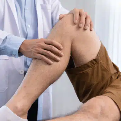
A meniscus transplant is a surgical procedure that replaces a damaged or missing meniscus with a meniscus allograft from a tissue donor. The donor meniscus must match your knee's size and shape to ensure proper outcomes.
The meniscus plays a vital role in knee biomechanics. It distributes weight across your joint and maintains joint space. Without it, your bones rub together and cause pain.
The procedure works through arthroscopy in most cases. Surgeons make small cuts and use a camera to guide the graft placement. Some cases need open surgery, especially when bone plug fixation is required for secure attachment.
This transplantation approach focuses on saving your natural knee. The allograft restores your knee's shock absorber function and protects cartilage health. Joint preservation delays the need for total knee replacement by years or decades.
The donor meniscus acts like your original tissue. It helps with load transmission across the joint. This reduces stress and protects against cartilage wear.
Proper patient selection ensures the best outcomes. Your surgeon evaluates your joint anatomy and activity level during patient evaluation. The goal is matching the right candidates with this joint preservation option.
The surgery begins with careful imaging and sizing of the allograft. Your doctor reviews MRI scans to measure your knee precisely. This ensures the graft fits perfectly.
During the procedure, surgeons prepare your knee joint. They remove any damaged tissue and create space for the new donor meniscus. The graft is then carefully positioned.
Fixation methods use sutures to attach the allograft to surrounding areas. Some techniques include bone plugs for stronger connection. The sutures hold everything in place during healing.
Blood supply to the outer edges helps integration. Your body gradually accepts the donor tissue. This healing process takes several months as the graft becomes part of your knee.
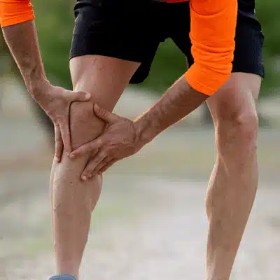
Patient selection determines transplant success and outcomes. Ideal candidates are younger than 50 and physically active. You need healthy cartilage in most of your knee.
Eligibility criteria include stable ligaments and good cartilage health. Your activity level should justify the surgery. Patients who want to return to sports often seek this treatment option.
Indications for surgery include pain after meniscus removal and early cartilage damage. The procedure works best when osteoarthritis hasn't advanced too far. You should have minimal cartilage wear in most areas.
Indications also include those experiencing knee injuries that led to meniscus loss. If you've tried other treatments without relief, transplantation may help. Your surgeon assesses whether joint preservation surgery fits your situation.
Contraindications exist for some patients. Advanced arthritis makes the procedure less effective. Unstable joints need other repairs first before considering a meniscus allograft.
Rehabilitation starts immediately after your procedure. You'll use crutches for several weeks to protect the graft. This allows proper healing without too much stress.
Physical therapy begins with gentle range of motion exercises. Your therapist gradually adds strength training. Full recovery takes six to twelve months for most patients.
Weight-bearing restrictions last about six weeks. Your medical team monitors the healing process with regular checkups. Following your rehabilitation plan improves outcomes significantly.
Return to sports requires medical clearance. Most patients resume full activities within a year. Your dedication to therapy determines your final outcomes.
Like any surgery, meniscus transplants carry risks that affect outcomes. Infection can occur but remains rare with proper care. Graft failure happens in some cases if the tissue doesn't heal correctly.
Blood clots are a potential complication your team will discuss. Stiffness might develop without proper therapy and rehabilitation. Joint lubrication problems can persist in rare situations.
The graft may not entirely integrate into your knee. Some patients require further operations to resolve difficulties. These issues can have long-term consequences.
Many dangers can be reduced via careful fixation during surgery. Following post-surgical advice is critical for avoiding complications. When patients strictly adhere to medical guidance, the majority of them achieve positive results.
Understanding the long-term benefits of joint preservation surgery
The allograft replaces your missing meniscus and brings back normal knee function. Your joint moves the way it was designed to move.
The new meniscus shields your cartilage from damage and wear. This protection keeps your knee healthy for years longer.
A working meniscus spreads weight evenly across your knee. Your bones receive better shock absorption and less stress.
The transplant may prevent or delay arthritis for many years. You avoid early joint replacement and maintain mobility.
Joint preservation is the main goal of this transplantation. The allograft restores your knee's natural biomechanics and function. It protects against further cartilage damage and wear.
The transplant may delay or prevent osteoarthritis for many years. Studies show improved outcomes in knee stability and mobility. Pain relief occurs as the joint stabilizes with the new tissue.
Cartilage restoration happens indirectly through better protection. The graft shields existing cartilage from excessive stress. This preservation approach beats early joint replacement for younger patients.
Joint preservation through meniscus transplant offers hope for active people. The procedure maintains your natural anatomy longer. Better outcomes occur when surgery happens before severe damage develops.
When facing knee injuries, you have several treatment options available. Meniscus repair works if enough healthy tissue remains in your knee. Removal relieves pain but causes outcomes like problems later with cartilage health.
Living without a meniscus leads to progressive cartilage wear. Total knee replacement is too extreme for young, active patients. A meniscus allograft fills the gap between these approaches.
Some patients combine procedures for better outcomes. Cartilage restoration techniques may accompany transplantation when needed. Your surgeon will recommend the best path based on indications and your specific knee condition.
The transplantation procedure offers a middle ground. It preserves your natural joint longer than other options. This knee preservation surgery delays more invasive procedures like total replacement.

Don't wait until your cartilage health declines further. If you've had a meniscus removed or severely damaged, talk to an orthopedic surgeon about meniscus transplant surgery today. Early intervention leads to better outcomes and can save your joint from premature arthritis.
Schedule a consultation to learn if you're a candidate for this procedure. Bring your imaging results and surgery records to your appointment. A specialist can perform a thorough evaluation and discuss whether this joint preservation option fits your needs and goals.
A donor meniscus can last 10 to 20 years or longer with proper care and rehabilitation. Success depends on factors like your age, graft fixation quality, and how well you follow recovery protocols.
Most patients return to sports within 9 to 12 months after the procedure. Your surgeon will clear you based on how the graft healed and your strength during therapy sessions.
Good candidates are under 50 years old with pain from meniscus loss but minimal arthritis. You need stable ligaments, healthy tissue in most areas, and realistic expectations about outcomes.
Surgeons use MRI scans and special imaging to measure your joint anatomy precisely. The tissue bank provides an allograft that matches your size within millimeters to ensure proper function.
Shoulder pain affects millions of people each year. Most cases improve with rest and simple treatments. But some problems need surgery to restore function and relieve discomfort.
This article explains when surgery becomes necessary. You'll learn the warning signs to watch for and what conditions require an operation. We'll also cover how doctors perform an evaluation and what treatment options are available.

Shoulder pain has many causes. Overuse injuries from sports or work are common. Age-related wear and tear plays a role too. Sudden trauma like a fall can damage the joint.
Common shoulder problems include tendinitis, bursitis, and arthritis. Tendinitis happens when tendons become inflamed from repetitive motion. Bursitis occurs when the fluid-filled sac in your shoulder gets irritated. Osteoarthritis breaks down cartilage over time.
Most shoulder issues like tendinitis don't require shoulder surgery. Only a small number of patients need an operation. Many conditions improve with conservative treatment like rest, medications, and physical therapy.
But some problems are more serious. They may not respond to basic care. The question becomes whether your symptoms are severe enough to consider surgery.
Several symptoms suggest you may need shoulder surgery. Pay attention to these key indicators.
Pain that won't stop: If shoulder pain lasts for months without improvement, that's a warning sign. Pain that disrupts your sleep or daily activities signals a serious issue. When physical therapy and rest don't help after three to six months, surgery may be your best option.
Limited movement: Struggling to lift your arm or reach overhead indicates joint damage. Stiffness that prevents basic tasks like getting dressed suggests surgery might help. Loss of range of motion from frozen shoulder or advanced arthritis often causes this problem.
Frequent dislocations: Shoulders that pop out repeatedly need attention. Shoulder instability leads to more damage over time. If your joint feels unstable or you fear it will dislocate, you likely need surgical repair. Frequent dislocation creates shoulder instability that requires fixing.
Significant weakness: Loss of shoulder strength points to tendon damage. Rotator cuff tears often cause this symptom. Large tears require rotator cuff repair to restore function. If you can't lift objects or raise your arm, surgery may be necessary.
Advanced joint damage: Severe arthritis that causes bone-on-bone grinding needs treatment. When medications and injections stop working, shoulder replacement becomes an option. This surgery can dramatically reduce pain and improve motion.
Injuries that won't heal: A fracture or major ligament tear sometimes needs surgical fixing. Sports injuries that don't respond to care may require an operation. If you're not healing after months of treatment, surgery may be required.
Your age, health, and lifestyle also matter. If symptoms limit your work or hobbies significantly, that's another warning sign. The decision depends on how much your shoulder affects your quality of life.

Doctors ensure you've tried other treatments before recommending surgery. During a consultation, an orthopedic specialist checks your symptoms carefully. They perform an examination of your range of motion, strength, and any swelling.
Imaging tests help confirm the diagnosis. X-rays show bone problems like a fracture or arthritis. An MRI reveals soft tissue damage like rotator cuff tears or severe tendinitis. Ultrasound can show inflammation in tendons or bursa.
Most doctors recommend trying conservative treatment first. Common approaches include rest, physical therapy, medications, and injections.
Physical therapy strengthens your shoulder and improves flexibility. Exercise programs often resolve issues like shoulder impingement or mild tendinitis. Anti-inflammatory medications reduce pain and inflammation. A corticosteroid injection provides temporary relief by reducing swelling in cases of bursitis or tendinitis.
The goal is to achieve pain relief without surgery risks. These treatments succeed in many cases. Doctors have you continue this care until it's clear symptoms aren't improving. If you've tried everything for a reasonable period and still hurt, it's time to discuss surgery.
The type of operation depends on your specific problem. Shoulder surgeries range from minimally invasive repairs to full joint replacement.
Arthroscopy: Many issues can be fixed with arthroscopy. The surgeon makes small cuts and uses a tiny camera. This approach treats rotator cuff tears, tendinitis, and shoulder instability. For chronic bursitis or impingement, the surgeon removes inflamed tissue and shaves bone. This creates more space and reduces pain. Arthroscopy typically offers faster recovery than open procedures.
Open repairs: Some complex problems need traditional open surgery. Large fractures may require plates and screws. Extensive tendon tears sometimes need direct access through larger cuts.
Shoulder replacement: End-stage joint damage requires replacing the joint. Total shoulder replacement uses prosthetic parts for the ball and socket. This helps severe osteoarthritis or rheumatoid arthritis. For massive, irreparable tears with arthritis, a reverse replacement may be used. This design lets other muscles power your shoulder. Shoulder replacement has high success rates for pain relief. It's becoming more common as implants improve.
Most arthroscopy is outpatient while shoulder replacement may need a short hospital stay.
A simple timeline and risk checklist to help set realistic expectations.
Wear sling for comfort, begin gentle assisted movements, manage pain with medications.
Start physical therapy, gradually increase range of motion, wean off strong pain meds.
Resume daily activities, begin strengthening exercises, 80–90% recovered.
Continue improving strength, return to sports and heavy activities, full recovery.
All surgery carries risks. But the benefits can be life-changing when done for the right reasons.
Benefits: The main advantage is lasting pain relief. Many patients report their pain completely disappears. Surgery also restores function so you can raise your arm and regain strength. You'll likely need fewer pain medications afterward.
Risks: Complications can include infections, bleeding, or blood clots. Nerve or blood vessel injury may cause numbness or weakness. Some patients develop stiffness or scar tissue. For replacements, joints can dislocate or loosen over years. Rotator cuff repairs might not heal fully or could tear again.
Despite these risks, serious complications are rare. Most patients feel the improvement is worth it.
Recovery: Healing takes time. Arthroscopy has shorter recovery periods. After major repairs, your arm stays in a sling for weeks. Full recovery can take months.
Rehabilitation through physical therapy is critical. You'll start with gentle movements and progress to strengthening. It often takes several months to achieve the best outcome. Most patients resume daily activities within weeks but avoid heavy lifting for longer.
By three to four months, many people are mostly recovered. Full strength may continue improving for up to a year.

Deciding on shoulder surgery is personal. Consult with a trusted orthopedic surgeon about your treatment options. Surgery is usually your choice based on when pain becomes unbearable or stops you from living normally.
The good news is that techniques keep advancing. In 2025, surgeons use refined methods and better implants. Success rates are high for restoring shoulder function.
Feel free to get a second opinion if you're unsure about alternatives. Clear information about your condition helps you make an informed decision. Surgery is never the first step. It's considered after other treatments fail or when damage is significant.
Take action now. If you have persistent shoulder pain that won't improve, don't wait. Schedule an evaluation with an orthopedic specialist to discuss your options. Early diagnosis and proper care prevent further damage and get you back to your normal life faster.
Most doctors recommend at least three to six months of physical therapy and other treatments. If your symptoms don't improve during this period, it may be time to discuss surgery. Severe injuries causing sudden weakness may need earlier intervention.
Many patients experience significant pain relief after surgery. However, some people may still have mild discomfort or limited motion. Results depend on the severity of your condition and how well you follow your recovery plan.
The top reasons include rotator cuff tears that don't heal, severe arthritis causing bone-on-bone grinding, frequent shoulder dislocations, and fractures that won't heal properly. Sports injuries and chronic tendinitis that don't respond to treatment also lead to surgery.
Recovery time varies by procedure type. Arthroscopy patients often return to light activities within weeks. Major repairs like rotator cuff surgery or shoulder replacement require several months. Most people continue improving for up to a year after surgery.
Surgery can feel scary. But getting the right information ahead of time makes a huge difference. Patient education means learning about your health, treatment options, and how to care for yourself before and after surgery. In 2025, giving patients clear health education has become standard in orthopedics and other fields. Research shows that informed patients get better health outcomes and feel happier with their care.
When you understand your condition and what surgery involves, you can make smart health decisions. You become part of your own care team. The benefits of patient education reach far beyond the operating room. They affect your recovery, your safety, and your quality of life.

One major win from pre-surgery education is improved health literacy. This means you can better grasp health information and follow instructions. Low health literacy levels have been linked to worse results after surgery. Patients with very low patient health literacy face 4.5 times higher odds of infection than those who can comprehend their care plans.
Good patient education materials raise your knowledge about the procedure, risks, and recovery steps. The American College of Surgeons found that preoperative health education and good patient education resources manages expectations and creates major positive effects on treatment outcomes.
The patient education process often uses different formats. Your care team might offer verbal counseling, printed patient education materials, videos, and online modules. When these patient education resources use plain language, they work better. You can weigh your options and join in health care decision crafting. The treatment plans align with your values and health care goals. Better-informed patients feel more confident and report higher patient satisfaction.
Facing surgery often brings anxiety and fear. Good health education can ease these feelings by replacing fear of the unknown with real knowledge. Research shows that informed patients feel less anxiety, develop better coping skills, and report higher patient satisfaction with their care.
When you learn what to expect, you go into surgery mentally ready. One study found that patients who got thorough pre-surgery education felt more in control. They had less discomfort and stress afterward. This led to quicker recovery and shorter hospital stays. Educating patients ahead of time turns an overwhelming experience into something manageable. It improves your care experience and emotional health.
An educated patient is an engaged patient. When you understand your condition and treatment, you take an active role instead of sitting on the sidelines. This increased patient engagement creates better communication with your healthcare team. You ask questions, share concerns, and work together on decisions. Better communication helps ensure everyone is on the same page about your care.
Patient empowerment happens when you feel ownership of your health journey. Giving a patient engagement with knowledge and control leads to better patient outcomes. In surgery, empowered patients might double-check important details. They become extra safety partners. This patient involvement improves both safety and results.
As patient engagement rises, so does patient satisfaction. You feel heard, respected, and involved. Patient activation means you become a proactive participant instead of a bystander.

Another major benefit is better treatment adherence and treatment compliance. When you fully understand your treatment plans and why each step matters, you follow them correctly.
Evidence shows that patient education interventions boost medication adherence. One analysis found that interventions improving communication at hospital discharge led to higher treatment adherence. These also significantly lowered readmission rates. When you know how and when to take your medications, you follow through.
Educated patients attend follow-up appointments and therapy sessions because they see these as key to recovery. These patient education resources help you adopt health behaviors that support healing. Health education helps you understand the reasons behind each recommendation. You get more consistent self-care, better pain control, and smoother healing. It also helps with disease management for any other health conditions you have.
The most compelling advantages are the real improvements in health outcomes. Many studies show that educating patients upfront leads to better results after surgery.
Patients who join patient education programs tend to recover faster and go home sooner. Several trials found that those receiving education had significantly shorter hospital stays than those with standard care. These improved health outcomes show why education matters so much.
Understanding how to care for yourself after surgery prevents many issues. Poor education contributes to hospital readmissions. Many readmissions happen simply because patients don't fully understand their conditions. By ensuring you have the knowledge to manage your health at home, hospitals have seen drops in readmission rates.
Educating patients also boosts patient safety. When you know about safety procedures, you can actively cooperate and even speak up if something seems wrong. An informed patient is more likely to mention important medical history details that could prevent medication errors.
How Patient Education Drives Long-Term Health
Smoking cessation before surgery leads to far fewer complications and faster healing
Losing weight before surgery reduces risks and improves recovery outcomes
Diet counseling improves nutrition and aids tissue healing after surgery
Patients continue healthy behaviors long after the operation for better fitness
Effective education saves $5.7M per year in postoperative care costs
Education promotes health prevention measures and wellness for life
The benefits often extend beyond immediate recovery. Educated patients are more likely to adopt health prevention measures and lifestyle changes that support long-term wellness. A preoperative program might encourage you to quit smoking or lose weight before surgery. Patients who get diet counseling might improve their nutrition. Those taught about exercise may continue those lifestyle changes long after the operation.
One analysis estimated that effective preoperative education could save roughly $5.7 million per year in postoperative care costs. This shows its impact on health care utilization and healthcare expenditures.
Healthcare providers now prioritize patient education initiatives as a formal part of surgical care. Many hospitals have established programs that provide structured patient education opportunities to learn and ask questions well ahead of your surgery date. This patient education integration into standard care ensures that everyone gets the health information they need.
Modern approaches also use technology. Patient education platforms like interactive modules, patient portals, and smartphone apps deliver information in engaging formats. These digital patient education resources reinforce what you learn in person and let you review materials at your own pace. More patient education resources mean more ways to learn in the way that works best for you.
To make education truly effective, healthcare teams must address patient empowerment obstacles. Common barriers include language differences, cultural beliefs, or too much information at once. Clinicians must listen to patient concerns and encourage questions. This approach might include discussing results of health risk assessments like blood sugar control.
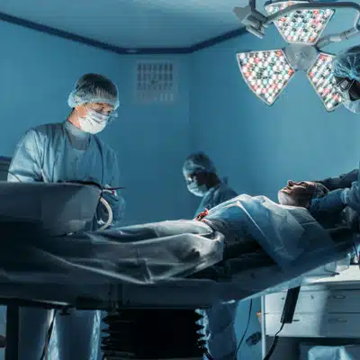
Educating patients before surgery is a powerful clinical tool. It improves outcomes, safety, and patient well-being. From boosting health literacy and confidence to reducing anxiety and problems, the benefits are proven across studies.
For healthcare systems, these improvements mean lower healthcare costs due to more efficient care. In 2025 and beyond, patient education is becoming an essential part of quality surgical care.
If you're scheduled for surgery, take action now. Enroll in any available programs your provider offers. Use all the patient education resources at your disposal. Ask questions. Review brochures, websites, and videos. In doing so, you take an active step toward safer surgery and better health. An informed patient is a safer patient. By partnering with your surgical team through education, you set the stage for a smoother operation, a faster recovery, and a healthier future.
How does patient education reduce complications after surgery?
Patient education teaches you how to care for yourself properly at home. You learn to spot warning signs of infection, follow wound care instructions, and know when to call your doctor.
What types of materials are most effective?
The best approach combines multiple formats to match different learning styles. Written handouts, videos, interactive online modules, and one-on-one conversations with your care team all work together.
Can patient education really reduce anxiety about surgery?
Yes, research consistently shows that informed patients experience less anxiety and fear. When you know what to expect at each stage, the unknown becomes manageable.
How early should I start learning about my upcoming surgery?
Start as soon as your surgery is scheduled. Early education gives you time to ask questions, make necessary changes like quitting smoking, and prepare mentally and physically.
Sports injuries don't have to sideline your athletic dreams. With the right approach, you can stay healthy while pushing your limits. Sports medicine research shows that smart prevention strategies can cut injury rates dramatically.

Strong bodies resist injury better than weak ones. Preseason conditioning creates this foundation before competition begins. Athletes who complete more preseason conditioning face fewer injuries during their season.
Your preseason conditioning program should start 6-8 weeks before competition. Begin slowly and build intensity week by week. This gradual approach lets your body adapt safely through proper training practices.
Strength training forms the core of injury prevention. Strong muscles protect joints and absorb impact forces. Research in sports medicine proves that muscle strength programs cut overall injuries by two-thirds. Overuse injuries drop by half.
Focus on exercises that build power throughout your body. Include squats, deadlifts, and lunges for lower body strength. Add push-ups, pull-ups, and rows for upper body power. Don't forget core strength work like planks and bridges. A strong core improves balance and body control.
Balance exercises deserve special attention in your preseason conditioning routine. These exercises train proprioception - your body's ability to sense its position. Better proprioception prevents ankle sprains and knee injuries. Studies show balance exercises alone reduce ankle injuries by 40%.
Add plyometrics to your preseason conditioning twice per week. These explosive jumping exercises build power and teach proper landing mechanics. Soccer players who added plyometrics to their preseason conditioning cut injury rates by over 50%.
Make your preseason conditioning sport-specific when possible. Runners should gradually increase mileage in their preseason conditioning programs. Basketball players need lateral movement drills in their preseason conditioning. Follow the 10% rule - never increase training load by more than 10% per week.
Balance exercises should include single-leg stands, wobble board training, and dynamic stability work. These prevention strategies target proprioception and reduce injury risk. Include balance exercises in every preseason conditioning session for best results.

Cold muscles tear easily. A proper warm up prepares your body for intense activity. Spend 5-10 minutes on light movement before practice or games. Jog, do jumping jacks, or cycle at an easy pace.
Follow your warm up with dynamic movements. Leg swings, arm circles, and walking lunges work well. These movements increase blood flow and improve flexibility. Flexible muscles resist injury better than tight ones.
Save static stretching for after your warm up. Hold each stretch for 20-30 seconds without bouncing. Focus on the muscle groups your sport uses most. Volleyball players should focus on shoulder flexibility. Runners need flexible calves, quads, and hip flexors.
Regular stretching improves joint range of motion over time. This makes injury prevention easier as the season progresses. Many exercise-related injuries happen when muscles lack adequate flexibility through poor stretching habits.
Your protective equipment serves as your last line of defense. Helmets prevent head injuries in football, hockey, and cycling. Shin guards protect soccer players from kicks. Mouthguards shield teeth in contact sports.
Modern protective equipment uses advanced materials to absorb impact. These designs reduce injury risk when worn correctly. The key word is "correctly" - poorly fitting gear won't protect you fully.
Check your safety gear regularly for damage or wear. Replace broken items immediately. Many helmet standards require yearly reconditioning to maintain protection levels. Never compromise on protective equipment quality or fit.
Don't overlook simple gear like proper footwear. Basketball shoes with ankle support help prevent sprains. Cleats give field sport athletes the traction they need. The right shoes for your sport and surface make a real difference.
Youth sports leagues should enforce equipment rules strictly. Coaches must do regular gear checks. Young athletes need extra education about why gear matters for their safety.

Good proper technique prevents injuries just as much as it improves performance. Football players who tackle with correct form avoid dangerous head and neck injuries. Soccer players learn safe heading technique once they reach the right age.
Coaching plays a huge role in injury prevention. Coaches must prioritize safety education alongside skill development. They should correct poor form immediately, both in games and practice.
Recent rule changes in youth sports show how policy can improve safety. When U.S. Soccer banned heading for players under 10, concussion rates dropped 26%. These prevention strategies work at the system level.
Education remains the foundation of all injury prevention efforts. Coaches need training in safety practices and injury recognition. Research shows that formal coach education leads to better adoption of safety measures. Sports education programs help coaches understand injury patterns and risk factors better.
Athletes must learn to communicate about pain and discomfort through proper education. A culture of openness prevents minor issues from becoming major injuries. Young players especially need education about listening to their bodies.
Take advantage of available safety resources through continuing education. The CDC offers concussion education programs. Sports medicine clinics provide injury prevention workshops. The more education you have, the safer you'll be.
Education should cover proper movement patterns, equipment use, and warning signs of injury. Ongoing education helps athletes and coaches stay current with best practices. Make education a priority in your sport participation.
Rest prevents injuries just as much as training does. About 50% of youth sports injuries come from overuse rather than acute trauma. These develop when athletes repeat the same motions without enough recovery time.
Take at least 1-2 complete rest days each week. Youth athletes should take 2-3 months off per year from their main sport. These breaks prevent injuries and mental burnout through proper rest periods.
Cross-training helps prevent repetitive stress injuries. Mix running, swimming, cycling, and other activities through agility training variations. Playing multiple sports through childhood actually reduces injury risk compared to early specialization.
Watch for signs of overtraining syndrome. This happens when athletes train too hard without enough recovery. Symptoms include chronic fatigue, declining performance, and increased injury risk.
Sleep plays a crucial role in muscle repair and growth. Growing athletes need extra sleep for tissue healing. Poor sleep increases injury risk and slows the recovery process.
Don't ignore pain signals from your body. Address aches and pains early before they become major problems requiring rehabilitation.

Good nutrition supports injury prevention and tissue repair. Protein helps rebuild damaged muscle tissue. Carbohydrates refuel energy stores after training. Calcium and vitamin D keep bones strong.
Hydration affects performance and safety directly. Dehydrated athletes face higher risks of muscle cramps, heat illness, and slower reaction times. Drink water before, during, and after exercise. Aim for one cup every 15-20 minutes in hot conditions.
If you do get injured, complete your rehabilitation fully before returning to play. Many athletes rush back too soon and get hurt again. Proper rehabilitation restores strength, flexibility, and proprioception through structured programs.
Work with a physical therapist or athletic trainer when possible. They can guide you through a program that addresses all aspects of healing. Think of rehabilitation as another form of injury prevention.
Sports injury prevention requires commitment from athletes, coaches, and parents. Start with comprehensive preseason conditioning that includes strength work, balance exercises, and sport-specific preparation. Your preseason conditioning program should address all movement patterns your sport requires.
Always warm up properly and maintain joint mobility through regular flexibility work. Use appropriate gear and learn safe techniques from qualified instructors. Prioritize adequate rest to prevent overuse problems.
Support your body with good nutrition and proper hydration. Complete any rehabilitation programs fully before returning to competition.
Most importantly, never stop learning about injury prevention through ongoing education. Knowledge continues advancing in this field. Stay informed through credible sources and safety programs.
At Sunnyvale Orthopedics, our specialists help athletes stay healthy and perform their best. We offer evaluations, prevention guidance, and expert treatment when needed.
Ready to prevent injuries and improve your performance? Contact Sunnyvale Orthopedics today to schedule your sports medicine consultation and take the first step toward safer athletics.
Shoulder pain affects up to 66% of people during their lifetime. With over 11 million specialist visits each year in the U.S., finding the right specialist matters. Your shoulder is a complex ball-and-socket joint with many muscles, tendons, and ligaments, and even a small cartilage tear can upset the joint. When pain strikes, you need an expert who understands this intricate system.
A shoulder pain specialist typically works in orthopedics or sports medicine, with rheumatology input when inflammation is suspected. They focus on shoulder conditions and have advanced training to help you heal, and may include massage therapy when it fits your plan. But how do you pick one you can trust? Here's what to look for when choosing your shoulder care provider.

Your shoulder is one of the most mobile joint systems in your body. This flexibility makes it prone to many problems. Common issues include rotator cuff tears, tendonitis, bursitis, impingement, arthritis, and trauma like dislocation or fractures.
Minor strains might heal with rest, ice, and NSAIDs. Ice works best for new injuries to reduce pain and inflammation. Heat helps later when muscles are tight and you want better range of motion.
But if pain lasts more than a few days or limits function, see a specialist. Waiting too long can lead to mobility issues or chronic problems that are harder to fix.
A good specialist can tell if your pain comes from a cartilage tear, nerve compression, or damage to the shoulder socket. They use proper diagnosis to guide your treatment plan. The right imaging helps confirm what's wrong. X-rays show bone problems. An ultrasound reveals how soft tissues move. An MRI scan displays all internal structures in detail.
Look for board certification in orthopedic surgery or sports medicine. Fellowship training in shoulder surgery shows extra commitment to quality care. The more often a specialist does a procedure, the better outcomes tend to be.
Ask how many years they've practiced and how often they handle cases like yours. If you might need shoulder replacement surgery or arthroscopy, find out their success rates. A trustworthy specialist will share this information openly.
Check if they belong to professional groups or do research. The FAAOS designation shows ongoing education. Get recommendations from your primary specialist or physical therapist. Read patient reviews, but use trusted sources and consider the context.
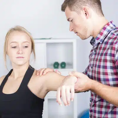
The best specialists offer many treatment options, not just surgery. They start with conservative care when possible. Rest, ice, and physical therapy often help with many shoulder injuries. Only severe cases need surgery right away.
Physical therapy is usually the first step. Guided strengthening exercises and stretches help rebuild shoulder strength and range of motion. A good specialist either has therapists on staff or works closely with experienced ones for your rehabilitation.
Your specialist might suggest other treatments too. Cortisone injections can reduce inflammation and provide short-term pain relief for severe flare-ups. They should explain these are temporary fixes, not long-term solutions.
Some clinics offer newer options like PRP therapy or stem cell therapy for certain injuries. These aim to help tendons and cartilage heal naturally. A current specialist will discuss if these fit your case.
Many patients find massage therapy helpful alongside medical care. Research shows massage therapy works as well as other active treatments for shoulder comfort and function. Acupuncture can also help with pain and muscle tension. A good specialist recognizes these complementary approaches in a complete shoulder treatment plan.
When conservative care isn't enough, your specialist should explain surgical options clearly. Many shoulder surgeries today use arthroscopy with small cuts and a tiny camera. This means less tissue damage and faster healing than open surgery.
For severe damage, you might need more extensive procedures. This could include shoulder replacement surgery or tendon transfers. A trusted specialist explains why they recommend a procedure, what it involves, and what to expect. They only suggest surgery when benefits clearly outweigh risks.

Technical skills aren't everything. You want a specialist who listens to your concerns and includes you in decisions. Great communication is vital. Your specialist should explain your diagnosis and treatments in plain terms. They should also hear your goals and worries.
Pay attention during your first visit. Does the team treat you well? Do they take time to check your shoulder thoroughly? A caring specialist acknowledges your pain and keeps you comfortable throughout the process.
Trust your gut feelings. If you feel at ease with the specialist and staff, that's a good sign. If the physician seems rushed or pushes treatment without explanation, consider getting a second opinion. A positive relationship can reduce anxiety and improve your recovery process.
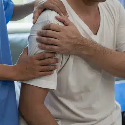
Start with recommendations from people you trust. Your primary specialist, friends, or family might know excellent specialists. Personal referrals often lead to specialists known for quality care.
Research the clinic or hospital. High-quality institutions usually have high standards for their orthopedic departments. Look for places that offer complete care including imaging, rehabilitation, and pain management all in one location.
Check what services they provide. A good specialist should handle everything from basic treatments to complex surgery. They might offer acupuncture, massage therapy, or other complementary treatments. Having continuity of care through all phases gives you peace of mind and better injury prevention guidance.
Consider practical matters too. Pick someone whose office is easy to reach. Shoulder injuries can limit driving, and you may need frequent visits for therapy.
Most importantly, choose someone you trust. Confidence builds when a specialist shows honesty, expertise, and real concern for your health. If you have doubts, get a second opinion. Any ethical specialist will support your right to do this.
Don't let shoulder pain control your life. The right specialist makes all the difference in your recovery. Look for proper credentials, experience with your condition, complete treatment options, and clear communication.
Your shoulder carries a lot throughout each day. Find someone strong to lean on for your care journey. Whether you're dealing with a labrum tear, chronic inflammation, or any other shoulder problem, expert help can restore your strength and freedom from pain.
Ready to find relief? Contact us today. Your path to better shoulder health starts with that first call.
Severe ankle pain from osteoarthritis can make simple tasks like walking feel impossible. When ankle arthritis pain won't respond to other treatments, surgery becomes the answer. Two main options exist: ankle replacement and ankle fusion (arthrodesis).
Both procedures can dramatically reduce pain and restore function. But they work in very different ways. One preserves movement while the other eliminates it completely. With guided physical therapy, many people see better quality of life, while doctors work to prevent complications.
The choice between ankle replacement and ankle fusion depends on your age, activity level, and specific condition. Understanding both options helps you make the best decision for your future. If your osteoarthritis is advanced, your surgeon will explain the tradeoffs.

Ankle replacement uses artificial parts to restore your ankle joint. The surgeon removes damaged cartilage and bone from your ankle. Then they place a metal prosthesis on your shinbone and ankle bone.
A plastic spacer sits between the metal parts. This acts like new cartilage, letting the joint surfaces glide smoothly. The artificial joint moves just like your natural ankle did before arthritis struck.
Modern technology has made ankle replacement much more reliable. About 90% of today's implants still work well after 10 years. The procedure preserves your ankle's range of motion and natural walking pattern.
This matters because keeping ankle mobility protects other joints. When your ankle can't move, your foot and knee must work harder. Over time, this extra stress can cause problems elsewhere.
Ankle fusion takes a completely different approach. Also called arthrodesis, this surgery permanently joins your shinbone to your ankle bone.
The surgeon removes all damaged cartilage from your ankle joint. They rough up the joint surfaces so bone can grow between them. Metal screws, plates, or bone grafts hold everything in place while healing occurs.
After several months, the bones fuse into one solid piece. Your ankle joint disappears completely. Without a joint, there's no more bone rubbing against bone. This eliminates arthritis pain.
Ankle fusion has been used for decades with excellent success rates. Once the bones heal together, pain relief is usually permanent. The trade-off is losing all ankle flexibility.

The biggest difference between these surgeries is what happens to your ankle joint afterward.
Ankle replacement preserves movement. Your new joint bends up and down like a healthy ankle. This allows normal walking on stairs, hills, and uneven ground. You maintain ankle function and joint mobility.
Ankle fusion eliminates movement. Your ankle becomes completely rigid. You lose flexibility but gain maximum stability. Walking requires other joints to compensate for the stiff ankle.
Both approaches provide excellent pain relief and improve quality of life. Studies show patient outcomes are similar between the two procedures. The key difference is how you move after surgery.
With ankle replacement, ankle biomechanics stay closer to normal. This reduces strain on nearby joints and protects long-term joint health. Fusion changes your walking pattern permanently, which may affect other areas over time.
Healing from ankle replacement and ankle fusion follows different paths.
Ankle replacement recovery typically involves 2-4 weeks without weight bearing. You'll wear a cast initially, then switch to a removable boot. Physical therapy starts early to restore strength and movement. Most people return to normal activities within 3-6 months.
Ankle fusion recovery takes longer upfront. You can't put weight on your foot for 6-8 weeks or more. The bones need time to grow together completely. Once healed, rehabilitation focuses on adapting to your new walking style rather than restoring movement.
Both procedures require patience during the recovery timelines. Following your surgeon's instructions prevents complications and ensures the best outcome.
Ankle replacement advantages:
Ankle fusion advantages:

Ankle replacement complications can include implant wear over time. Active patients may need a revision surgery after 10-15 years. The procedure requires good bone health for the implants to anchor properly.
Ankle fusion drawbacks center on activity limitations. You lose all ankle flexibility permanently. The altered gait can stress your foot and knee joints. Some patients develop arthritis in these areas years later.
Both procedures carry standard surgical risks like infection or healing problems. Choosing an experienced surgeon minimizes these concerns.
Several factors guide this important decision:
Age matters. Older patients often prefer ankle replacement to maintain mobility. Younger people may choose fusion for its permanent solution without future surgeries.
Activity level counts. Heavy laborers and athletes often get fusion for maximum durability. Less active people may benefit more from preserved movement.
Bone health is crucial. Strong bones support ankle replacement well. Poor bone quality may favor fusion with bone grafts.
Ankle condition plays a role. Severe ankle deformities sometimes require fusion for deformity correction. Well-aligned joints with isolated osteoarthritis often do better with replacement.
Your surgeon will consider all these mobility considerations during decision making. They'll also discuss your personal goals and preferences. Patient choice should be informed by understanding both treatment options completely.

Advances in surgical technology continue improving both procedures. New implant designs last longer and feel more natural. Surgical techniques become less invasive each year, supporting ankle function.
Joint lubrication in artificial ankles now mimics natural cartilage better. This reduces wear and extends implant life. Some patients get 15-20 years from modern ankle replacements.
Fusion techniques (arthrodesis) also improve with better hardware and bone grafting methods. Healing times decrease while success rates climb higher.
Both surgeries now offer excellent long-term outcomes when matched to the right patient.
Choosing between ankle replacement and ankle fusion requires careful consideration. Both procedures effectively treat severe osteoarthritis and restore function.
Schedule a consultation with an experienced foot and ankle specialist. They'll evaluate your specific condition, discuss your goals, and may recommend gait analysis to understand your needs better.
The right surgery depends on your unique situation. Age, activity level, bone health, and personal preferences all matter. Some patients prioritize movement while others value permanent stability.
Don't let ankle arthritis pain control your life any longer. Modern surgery offers reliable solutions to get you back on your feet. Whether through ankle replacement or ankle fusion, you can look forward to years of improved function and freedom from pain.
Contact a qualified orthopedic surgeon today to explore which option best fits your needs and lifestyle goals.
An ankle fracture means one or more bones in your ankle joint are broken. This common injury affects thousands of people each year. While some ankle fractures heal with a cast, others require surgery to fix the broken bones properly.
Understanding when you need ankle fracture surgery and what to expect can help you make informed decisions about your care.

Your ankle joint contains three bones. The tibia forms the inner side, the fibula creates the outer side, and the talus connects to your heel bone. Strong ankle ligaments hold these bones together.
Ankle fractures happen when trauma damages one or more of these bones. Common causes include sports injuries, falls, car accidents, or simply stepping wrong. The injury can range from a simple crack to multiple breaks with torn soft tissues.
Signs of a fractured ankle include sudden severe pain, swelling, and being unable to put weight on the injured ankle. You might also see bruising or notice your ankle looks deformed.
Doctors diagnose ankle fractures using X-rays. Sometimes they need CT scans or MRI to see all the damage to bones and soft tissues.
Different types of ankle fractures affect different bones:
Lateral malleolus fracture breaks the fibula on the outside of the ankle.
Medial malleolus fracture splits the tibia’s inner edge, often showing a painful bump and ankle deformity. Posterior malleolus fracture involves the back of the tibia and may scatter bone fragments.
Bimalleolar fracture shatters both outer and inner bones, while bimalleolar equivalent combines one break with ligament damage, leaving the joint equally unstable.
Trimalleolar fracture injures all three malleolus fractures, demanding open reduction with hardware under anesthesia, usually at an outpatient surgery center.
Early pain management, careful weight bearing guidance, and physical therapy restore mobility and healing after fixation.

Not every broken ankle needs surgery; small fractures that stay often heal in a cast. Surgery is advised when bone pieces are displaced, multiple bones break, the fracture enters the joint, or ligament damage creates ankle instability. Open fractures also require urgent clean out and fixation.
Modern surgical techniques use small incisions to realign bones, fix a syndesmosis injury, and perform tendon repair when needed. Metal plates, hardware, and screws hold the ankle steady so weight bearing can resume safely during the healing process. Early motion exercises protect range of motion, while thinners reduce deep vein thrombosis risk and aid recovery.
Ankle fracture surgery relies on open reduction and internal fixation. After anesthesia the surgeon makes small incisions, exposes the break, and realigns the fragments.
Internal fixation follows: a plate and screws secure a lateral malleolus fracture along the fibula, while a medial malleolus break receives screws or a mini-plate. If ligaments are torn or a syndesmosis injury is present, a trans-syndesmotic screw holds the tibia and fibula together to protect bone healing.
In-surgery X-rays confirm alignment, then the wounds are closed and the ankle placed in a splint or walking boot. Complications are rare, and hardware usually remains permanently.

Healing from ankle surgery takes time and patience. Your recovery happens in stages:
First six weeks after anesthesia wears off, keep all weight off the repaired ankle. Use crutches or a knee scooter, elevate the leg above heart level, apply ice, and watch the incision for infection.
A splint or cast protects fixation, especially if arthroscopy, tendon repair, or syndesmosis injury complicated surgery.
At six to twelve weeks, X-rays confirm healing, then gradual weight bearing begins, often in a walking boot. Physical therapy minimizes scar tissue, restoring range, strength, and balance.
Most people walk normally by three or four months, though sports may wait six months and complex fractures can require a year.

Several steps help ensure successful recovery:
Follow weight-bearing rules strictly. Bearing weight too early can shift the break, especially with malleolus fractures, leading to poor healing.
Manage swelling and pain: elevate, ice, and take pain medication as prescribed. Keep the incision clean and dry, watch for infection, and attend every follow-up visit.
Prevent blood clots with ankle pumps, compression stockings, and prompt reporting of calf pain. Good nutrition with protein, calcium, and vitamin D speeds bone repair.
Do not smoke because it slows recovery. Stick to physical therapy to restore motion and strength.
These steps help patients with malleolus fractures or syndesmosis injury return to normal activities quickly.
Modern ankle fracture treatment offers excellent results for most patients. Surgical methods continue to improve, giving doctors better ways to fix broken bones and speed recovery.
The key is getting proper treatment quickly. Delayed care can lead to poor healing and long-term ankle pain. If you suspect an ankle fracture, seek medical attention right away.
Your orthopedic surgeon will explain all treatment options based on your specific injury. They consider factors like your age, activity level, and overall health when recommending surgery or other approaches.
Remember that recovery takes time, but most people regain full function of their ankle joint. With proper care and dedication to rehabilitation, you can expect to return to all the activities you enjoyed before your injury.
Don't let ankle pain control your