(214) 420-7611


Adhesive capsulitis, also called frozen shoulder, can occur when you are sidelined due to an injury or certain medical conditions. The resulting stiffness and pain can make everyday activities difficult.
Left untreated, a frozen shoulder can worsen and significantly interfere with daily life. So, what is this condition, and what is the most effective treatment for frozen shoulder?
A frozen shoulder is exactly what it seems like. Your shoulder becomes stuck, and mobility is limited as a result. You don't just don't function either; a frozen shoulder is normally painful.
In the shoulder joint, the upper arm bone's rounded shape fits into a socket in your shoulder blade. The joint is surrounded by connective tissue called the shoulder capsule.

Normally, the shoulder capsule effortlessly expands and contracts with arm movement. However, scarring develops when the tissues become inflamed, tightening the shoulder capsule, restricting movement, and causing pain. If you're in your situation, have a frozen shoulder.
A frozen shoulder is often directly related to a lack of movement. It often occurs in people who've had to keep their shoulder immobile after a broken arm, rotator cuff injury, or surgery and individuals who have conditions that limit their mobility, such as stroke or Parkinson's Disease. Reduced movement due to chronic tightness in your shoulder muscles contributes to an idiopathic frozen shoulder with no obvious trigger.
Our connective tissue adjusts based on how much we move – it tightens with less activity and loosens with more. This adjustment might set off a troublesome cycle: Moving becomes trickier when the joint capsule tightens and occasionally causes discomfort. This restriction hinders movement even more, prompting the tissues to tighten further.
Inflammation has an important role in developing a frozen shoulder as well. After surgery or injury, inflammation occurs in your joint as part of the healing process. This causes pain, swelling, and limited movement, resulting in scar tissue forming, making your joint capsule thicker and tighter.

Systemic inflammatory conditions, such as diabetes and other metabolic conditions, thyroid disease, osteoarthritis, cardiovascular disease, and autoimmune diseases, can also boost the risk of developing a frozen shoulder.
Most people with adhesive capsulitis experience worsening pain and a loss of movement. Adhesive capsulitis can be divided into four phases; your physical therapist can assist you to determine your stage.
During stage 1 of its development, it might be difficult to identify your issue as adhesive capsulitis. You've had symptoms for 1-3 months, and they're getting worse. Shoulder movement causes pain. It usually aches when you are not using it, but the pain increases and gets "sharp" with movement. You'll start to limit shoulder motion during this period and protect your shoulder by using it less.
The movement loss is most visible in "external rotation" (when you rotate your arm away from your body), but you may lose motion once you reach behind your back or raise your arm. Pain is the distinguishing feature of this stage; you might experience pain during the day and night.
At this stage, you've been experiencing symptoms for a period of 3 to 9 months, probably noticing a gradual reduction in shoulder movement and an uptick in pain, especially during nighttime. Although the shoulder retains some mobility, pain, and stiffness impose limitations on it.
Your symptoms have persisted for 9-14 months, and you have a greatly reduced range of shoulder movement. During the early part of this stage, there's still a substantial amount of pain. However, toward the end of this stage, pain decreases, typically occurring only when you move your shoulder as far as you can.
You've had symptoms for 12-15 months, and there is a huge decrease in pain, particularly at night. You still have a limited range of movement, but the ability to complete your day-to-day activities involving overhead motion is improving rapidly.

Often, physical therapists do not see patients with adhesive capsulitis until well into the freezing stage or early in the frozen phase. Sometimes, people are treated for other shoulder conditions once their physical therapist notices the signs and symptoms of adhesive capsulitis. Your physical therapist will thoroughly evaluate, including an extensive health history, to exclude other diagnoses.
Your physical therapist will search for a specific pattern in your decreased range of motion known as a "capsular "pattern" that is "typical with adhesive capsulitis. Moreover, your physical therapist will consider other conditions you might have, like thyroid disorders, diabetes, and autoimmune disorders associated with adhesive capsulitis.
The physical therapist's overall goal is to restore the movement so you can perform your day-to-day activities. Once the evaluation process has identified the stage of the condition, your physical therapist will create a tailored exercise program custom to your specific needs. Exercise is most effective for people who are in stage 2 or higher. Your treatment might include:
Exercises and manual therapy. The physical therapist will help you maintain as much range of motion as feasible and lessen the pain. Your therapist might use manual therapy (hands-on) techniques and range-of-motion exercises to maintain shoulder movement.
Modalities. Your physical therapist might use ice and heat treatments (modalities) to help relax your muscles before other forms of treatment.
Home-exercise program. The physical therapist will give you a gentle home exercise program to help reduce your loss of motion. The therapist will warn you that overly aggressive stretching in this phase may worsen your shoulder pain.
Your physical therapist will suit your treatment activities and intensity to the symptoms and inform you about the proper use of the affected arm. Your therapist will meticulously monitor your progress to ensure a safe healing procedure is observed.

Pain medication. Sometimes, conservative care can't reduce the pain of adhesive capsulitis. In that case, the physical therapist might refer you for an injection of a safe pain-relieving and anti-inflammatory medication. Research has shown that although these injections don't provide long-term benefits for range of motion and don't limit the duration of the condition, they do provide short-term pain reduction.
The focus of treatment during stage 3 is on the return of motion. Treatment might include:
In the final phase, your physical therapist will concentrate on the return of "normal" shoulder body mechanics and your return to normal, pain-free activities. Your treatment might include:
The reason for adhesive capsulitis is debatable, with no definitive cause. Hence, to date, there's no known method of prevention. The condition's onset is usually gradual, with the disease process having to "run its course." Nevertheless, the sooner you contact your physical therapist, the sooner you receive appropriate information on addressing your symptoms most effectively.
Joint replacement surgery is a significant medical procedure that has impacted many lives. It's a topic of interest for individuals suffering from joint pain and degenerative conditions, leading them to ask a pivotal question: "How successful is joint replacement surgery?"
This blog aims to provide clear, concise information about joint replacement surgery's success rates, processes, and considerations. It's crafted to be informative, straightforward, and helpful for those seeking a better understanding of what to expect from this medical intervention.

When considering joint replacement surgery, the primary concern is its success rate. How successful is joint replacement surgery? To answer this, it's essential to look at the outcomes experienced by patients. After this surgery, many patients experience significant pain relief and improved mobility.
Studies show that knee and hip replacements can have success rates as high as 90-95% over a 10-year period, which is quite promising.
However, success depends on various factors like the patient's health condition, the skill of the surgeon for joint replacement procedures, and post-operative care.
The definition of 'success' can vary from patient to patient - for some, it's the ability to perform daily activities without pain. In contrast, it might mean returning to more vigorous activities or sports for others.

A common question is whether joint replacement surgery can end arthritis. While the success of joint replacement surgery in relieving pain and improving function is well-documented, it's crucial to understand that this surgery doesn't 'cure' arthritis. What it does is replace the damaged joint surfaces with artificial components, which alleviates pain and improves joint function.
This means that while arthritis symptoms in the replaced joint are significantly reduced or eliminated, it doesn't prevent arthritis from possibly developing in other joints. Patients should follow their doctor's advice and maintain a healthy lifestyle to manage their joint health. This approach helps maximize the success of the surgery and enhances the overall quality of life.
Understanding the nature of joint replacement surgery is crucial for patients considering this option. Indeed, joint replacement is categorized as major surgery. This categorization is due to several factors, including the complexity of the procedure, the need for anesthesia, and the recovery time involved. But how successful is joint replacement surgery in terms of patient outcomes?
The success of joint replacement surgery is generally high, and many patients experience a significant improvement in their quality of life. The damaged joint surfaces are surgically removed and replaced with artificial ones. However, as with any major surgery, there are risks involved, such as infection, blood clots, or implant failure. Patients must discuss the risks with their healthcare provider before making a decision.
Joint replacement surgeries are most commonly performed on hips and knees, as these joints bear significant body weight and are prone to wear and tear. Hip and knee replacements are thus among the most successful medical procedures for improving a patient's life.
So, when addressing the question of how successful is joint replacement surgery, it's essential to note that these two types of surgeries have a long history of high success rates.
Other joints like shoulders, ankles, and elbows can also be replaced, but these are rare. The success of these surgeries, like hip and knee replacements, depends on various factors. This includes the patient's overall health, joint damage severity, and post-operative care and rehabilitation quality.
For those contemplating joint replacement surgery, exploring alternatives is a natural step. While pondering over how successful is joint replacement surgery, it's also important to consider other treatment options.
Alternatives to joint replacement can include nonsurgical methods like physical therapy, medications, lifestyle changes, and less invasive surgical procedures like arthroscopy.
Physical therapy and exercise can improve joint strength, flexibility, and pain. Medications, both over-the-counter and prescription, can help manage pain and inflammation.
Lifestyle modifications can help alleviate joint pain, including weight loss and avoiding aggravating activities. For some patients, arthroscopy, which is less invasive than joint replacement, can provide relief by repairing or cleaning the damaged joint.
It's important to note that while nonsurgical options can be effective, they may not provide the same long-term benefits as joint replacement surgery for specific conditions. Therefore, discussing with a healthcare professional is crucial for making an informed decision.
After joint replacement surgery, post-surgery care and rehabilitation are pivotal in determining the procedure's success. Addressing how successful is joint replacement surgery means also focusing on the recovery phase. Post-operative care involves managing pain, preventing infection, and starting physical therapy.
Rehabilitation is crucial in regaining strength and mobility in the affected joint. Physical therapy typically starts soon after surgery and lasts several weeks or months. The rehabilitation goals are to enhance joint function, expand range of motion, and facilitate return to daily activities. Patients are usually advised to follow a specific exercise regimen and may receive guidance on performing activities safely.

When discussing how successful is joint replacement surgery, it's imperative to look at the long-term outcomes. The majority of joint replacements have a lifespan of 15-20 years, with some lasting even longer. The success of any surgery depends on various factors such as age, activity level, and a healthy lifestyle.
Over time, joint replacements can wear out or become loose, potentially requiring revision surgery. However, with advancements in surgical techniques and implant materials, the durability of joint replacements has significantly improved. Patients need to have regular follow-ups with their orthopedic surgeon to monitor the condition of their joint replacement.
Addressing the pivotal question - how successful is joint replacement surgery - brings us to a reassuring answer. This surgery stands as a highly effective intervention for those struggling with severe joint pain and mobility issues. Advancements in medical technology and surgical procedures have made surgery safer and more successful, even though it remains a significant medical intervention.
Engaging with healthcare professionals who can provide comprehensive information and support is vital for anyone considering this path. A patient-centered approach, where the treatment is tailored to individual needs and goals, is crucial for the best outcomes. Joint replacement surgery relieves pain and empowers individuals to engage in daily activities and experience a more active and fulfilling lifestyle.
At Sunnyvale Sports Medicine and Orthopedic Center, we are dedicated to guiding our patients through their journey with expert care and support.
Many people wonder, "How long does a microfracture take to heal?" This is because this type of bone hurt tends to be on their minds. Microfractures, little but important bone breaks happen more often than you may think. They can come from extensive activities or even minor accidents. Unlike more significant breaks, their size doesn't make them less important in everyday life. It's important to know about this condition if you want help getting better.
We will look at not only how long it takes for these little breaks to get better but also what affects this job. If you're a sportsperson, weekend player, or someone who got hurt when they didn't expect it, this article is for helping and directing you to get better.
Small bone cracks, even though tiny, pose a big problem in the area of bone injuries. They happen when small breaks or cracks occur in the bone, usually because of too much use or constant stress. Imagine an athlete always jumping or a normal person with just a slight fall; these times can cause tiny cracks. These do not break the whole bone like complete fractures, but they can still make it hard to move and cause pain. The body's answer to microfracture is an interesting, complex process.
Initial Response: As soon as you get hurt, your body starts to work. Blood vessels at the hurt place break, causing a clot. This clot helps in making new bones grow.

Inflammation Phase: In just a few days, the spot gets bigger and starts to hurt. This swelling is how the body keeps damage safe and begins healing.
Bone Production: Special bone-making cells, named osteoblasts, start to create new bones. They work very hard, just like builders on a job site, making new bone material.
Remodeling Phase: This is the final touch. The new bone that has just formed is not strong at first and needs to get stronger. In a few weeks to months, the bone changes itself. It gets stronger and tougher.
The healing time for a microfracture largely depends on various factors, including the individual's health, age, and the location of the injury. Typically, the healing process can span anywhere from 6 to 12 weeks. This timeline, however, is not set in stone and can vary based on several considerations.
Factors influencing healing time include:
Pain from a microfracture can be very different. It could feel just a little bit uncomfortable, or it could hurt really badly, depending on how bad the injury is and what level of pain someone can handle. At first, the hurt can feel really strong and sharp. This is most common in the early days after getting injured. As you get better, the pain usually lessens.

Medication: Pills you can buy without a doctor's help, like acetaminophen or ibuprofen, usually lessen pain and swelling.
Rest and Elevation: Keep your hurt spot still and raised up to lessen pain from swelling.
Cold Therapy: Using cold bags on the first days after being hurt helps to lessen pain and reduce swelling.
Physical Therapy: When the bad pain goes away, light exercises help make stiffness less and improve how we move. This then helps to reduce discomfort.
Rest is very important for healing a microfracture. Taking good care and getting enough sleep not only helps you get better quickly, it also makes sure the healing works fully. But, it's hard to find a good sleeping position after having microfracture surgery.
Here are some strategies to ensure a comfortable night's sleep during recovery:
Elevate the Injured Area: Raising the hurt arm or leg can help lessen pain and puffiness. Use pillows to lift the hurt part slightly higher than your heart's level.
Use Supportive Bedding: A strong mattress and pillows can give the needed support. Think about using a body pillow or knee support for extra ease.
Find a Comfortable Position: Lying on your back while sleeping is usually suggested. If you hurt your leg or foot, avoid putting weight on it. If you have to sleep on your side, use a pillow between your knees for the right position and help.
Limit Movement: Try to reduce moving around a lot. If you have to switch places, be careful not to make big, fast moves that could make the hurt worse.
Prepare for Sleep: Make a relaxing bedtime routine. Don't drink coffee before going to bed, and try activities like reading or listening to gentle music that help you relax.
Everyone's body has a different way of responding when they get hurt. It might take a bit of time to find the best way to sleep. Ask your health doctor for advice made just for you. It's very important to get good sleep for healing, so make your sleeping place as comfy as you can.
When it comes to bone injuries, it's easy to get mixed up between a stress fracture and a microfracture.

Think of a stress fracture as a small but significant crack in the bone. It usually sneaks up on you, especially if you're someone who loves activities like long-distance running or basketball. You might start feeling a nagging pain in your lower leg or foot that just gets worse whenever you're on your feet. Healing from this takes about 6 to 8 weeks of laying low and giving your body a break from all that action.
Now, let's talk about a microfracture. It's a tinier break in the bone, more like a hairline crack, often caused by overdoing it or a minor bump or fall. Unlike stress fractures that prefer your legs and feet, microfractures can show up anywhere, like in your knees. The pain here can be sharp, and you might feel swelling or even a weird locking sensation in your joint. Healing time? It's a bit of a mixed bag, usually between 6 to 12 weeks, and it's all about keeping that joint as stress-free as possible.
So, while both are pesky bone issues caused by stress, they're different in where they strike, how they feel, and how you deal with them. Remember, if you're feeling off in the bones, it's best to chat with a doctor to get the real scoop on what's going on.
Healing from microfracture can feel like an adventure, but it won't take long if you do the right things. First things first: Do what the doctor said in his letter. They are experts and their advice is worth a lot. Rest is now your best friend. Let your body get the rest it needs to fix itself up.
Now, let's talk food. Eating right not only makes your taste buds happy, it's also good for your bones. Get some foods that are high in calcium and vitamin D. Remember food from cows, vegetables with leaves and some nice sunlight.
When it comes to prevention, everything is about balance. Change your exercise plan so you don't use the same bones and muscles all the time. Doing weight lifting can help a lot, it makes the muscles that keep those bones strong. Remember, listen to your body too. It's really smart and will tell you to slow down.
And there you have it – a full journey exploring "How Long Does a Microfracture Take to Heal". We've traveled from understanding what a microfracture really means, through the twists and turns of pain management, all the way to the steps for a strong recovery and smart prevention.
Remember, healing isn't a race; it's more like a leisurely stroll through the park. Each person's path is unique, with its own set of twists, turns, and rest stops. Embrace this journey with patience and a dollop of self-care. And hey, you're not walking this path alone. Sunnyvale Sports Medicine is here for you like a trusted guide, ready to support and steer you towards a safe and effective recovery.
When the menisci in your knees are damaged or degenerated, it leads to a torn meniscus. Athletes often experience this injury due to overuse, but torn menisci can also happen as you age, owing to the natural process of degeneration. This condition may need surgery, and many of our patients ask us, “Is meniscus surgery a major surgery?
Menisci, those crescent-shaped fibrocartilage pieces in your knee joint, play a crucial role in its well-being. You have a pair between your femur and tibia bones, assisting with the load-bearing and shock-absorbing duties.
Initially, a torn meniscus might not show symptoms. Yet, if you keep using the injured menisci, you could end up with persistent swelling, pain, and even some clicking sounds. Symptoms might get worse, especially post-activity.
Dealing with a torn meniscus offers various conservative options—rest, pain relief, and physical therapy. Your doctor might recommend surgery based on the tear's size and other health factors.

Let's discuss the various surgical choices for torn menisci, covering what to anticipate in recovery, potential issues, and the overall outlook for each surgery type.
Menisci can tear in various manners, and the tear kind can aid in deciding the optimal treatment approach:
Signs of a torn meniscus involve sharp knee joint pain, swelling, stiffness, and a sensation of "catching" during joint movement. Those with a torn meniscus may also feel a "popping" when the tear happens.
Your treatment depends on the kind and size of the tear and the damage around your knee. X-rays or MRIs (or both) and a physical joint check guide your treatment. Smaller meniscus and many aging tears often get better with rest, ice, anti-inflammatory meds, and sometimes injections into the joint to ease swelling and pain. Alternatively, a less invasive procedure, knee arthroscopy, might be a good choice.
Cartilage and the meniscus struggle to heal due to limited blood supply. Major tears usually can't fully heal; treatment focuses on cutting out the damaged parts to help "restabilize" the meniscus for firm joint support. This process, a meniscectomy, is done with special tools through small knee cuts. After surgery, you can usually move and bear weight on your knee soon.
When damage is on the outer rim with a better blood supply, special sutures or fasteners can be used to mend the meniscus, letting torn edges fuse during recovery. Meniscus repair takes longer to heal than a meniscectomy, and you might have weight-bearing restrictions, needing a knee brace in the early recovery stages.
Meniscus surgery is still one of the most common orthopedic procedures because many people have meniscus tears affecting their quality of life. If standard treatments don't work or the tear is severe, your doctor might suggest surgery to help you return to your usual activity level.
Surgery for a torn meniscus is not always the first choice. If you might have symptoms of a torn meniscus, your doctor might first recommend the "RICE" method:
You can reduce inflammation and prevent more damage by icing your knee and taking a break from sports or other intense activities. Alongside RICE, non-steroidal anti-inflammatory drugs (NSAIDs) like aspirin or ibuprofen may cut pain and swelling. Wearing a knee brace can also be beneficial.
Your doctor might also suggest a corticosteroid injection into the joint. Cortisone shots have been proven to relieve pain for patients with meniscal tears or degeneration substantially.
Yale Medicine explains that meniscus injuries in older people often result from degeneration over time. This can be caused by routine activities such as squatting or climbing stairs.

The steps involved in meniscal repair surgery include:
2-3 small (< 1cm) portals are created to allow passage of a camera and surgical tools within the knee joint. The whole joint is inspected for damage.
The torn meniscus is repaired using stitches. A repair is preferable to a resection as the shock-absorbing function of the meniscus is retained. Sometimes, this can be performed completely via keyhole surgery using an "all-inside" surgical device.
If the tear is extensive, an accessory incision is made on the side of the knee, and sutures are passed from the inside of the knee to the outside through the open accessory incision. These stitches are then directly tied over the capsule to provide strong repair. This is considered the gold standard for meniscal repair.
In some situations, the torn meniscus is unsuitable for repair, and the torn portion is trimmed to a stable margin. This can relieve the pain and mechanical symptoms associated with a tear. Trimming the meniscus will reduce its shock-absorbing capacity and put more pressure on the surrounding cartilage. This will cause the cartilage to wear out quicker than normal and lead to arthritis in the knee.

Individuals engaged in active office tasks or very light work with various responsibilities can resume work in approximately two or three weeks, contingent on lifting requirements and whether their employer is willing to adjust for lighter duties.
Tasks involving heavy labor (such as lifting or working at unprotected heights) typically can only be resumed after six weeks. Approval from their physical therapist is usually necessary for most cases. Engaging in the most strenuous labor, particularly working at unprotected heights, would naturally require a more extended recovery period.
Regarding driving, patients recovering from right knee surgery and left knee surgery (with a standard transmission car) should refrain from driving until they are out of the knee immobilizer, off all pain medications, and can fully bear weight without experiencing pain. Left knee surgery patients can resume driving after approximately three weeks if they are pain-free, off all pain medications, and can walk comfortably without crutches.

Injuries like a torn meniscus are quite common, especially among athletes and older individuals. It's crucial to get treatment to avoid more harm and deterioration quickly. Your doctor will probably suggest using over-the-counter pain relievers and starting with the RICE method, especially for mild cases.
But if your knee pain and swelling persist, disrupt your daily routine, or your tear is larger than 1 cm, you should talk to your doctor about surgery. This could be a partial meniscectomy or repair surgery.
Choosing surgery is a significant decision and comes with potential complications during recovery and later on. Before deciding on surgery for a torn meniscus, thoroughly review the advantages and risks with your doctor.
Whether bowling, rowing a canoe, lifting boxes, or pushing furniture, we rely heavily on our shoulders for several daily activities. Normally, the shoulder can move a lot, giving it the ability to be the most flexible joint in your body. However, because of this flexibility, it is not very stable and is prone to injuries.
Soft tissue refers to ligaments, muscles, and tendons surrounding and supporting our joints. A soft tissue injury affects the tendons, muscles, and ligaments. Soft tissue injuries to the shoulder can occur as a result of a fall on an outstretched hand, overuse, or direct trauma. Before we answer the most commonly asked question, “What are the most common shoulder soft tissue injuries?” let us first talk about the anatomy of the shoulder.
The shoulder is a ball-and-socket joint. The upper arm bone, is capped with a ball-like structure that fits the shoulder anatomy into a shallow, cup-shaped socket in the shoulder blade (scapula). The labrum, a strong cartilage ring that lines the socket, gives the ball of the humerus extra depth in which to nestle.

The joint capsule is a type of protective sleeve of tissue surrounding the joint and comprises part of the ligaments that link the bones and the tendons that comprise the rotator cuff.
Most sports injuries involving the shoulder damage the connective tissues: the labrum, ligaments, and tendons. Issues with these shoulder components are far more common than broken bones and bruised muscles due to the high-force, high-repetition activities required by athletics.
The rotator cuff is a set of four upper arm muscles. They let you raise and rotate your arm. Tendons connect the muscles to the bones. If the tendons rip, the humerus cannot move as freely in the socket, making it difficult to raise or lower the arm.
As we age and become less active, tendons degenerate and lose vigor. This can cause rotator cuff tears.
Rotator cuff tears are mostly seen among the middle-aged and elderly with pre-existing shoulder problems. As for younger people, overhead arm use, repetitive movements, and stress to the tendons can lead to rotator cuff injuries.
Shoulder sprain refers to a separated shoulder or acromioclavicular joint injury.
The A.C. joint is where the acromion (the bony projection at the top of your shoulder blade)meets the clavicle or collarbone. In shoulder sprain injuries, ligaments that support and stabilize the shoulders are stretched, the shoulder is stretched or torn, and the bones of the A.C. joint become dislocated or separated.
Trauma to the shoulder is the primary cause of shoulder sprain injuries, E.g., a car accident and falling onto an outstretched arm.

A shoulder tear occurs because of the injury to the soft tissues that provide the joint range of motion and stability. Tears can occur in the tendons, muscles, or labrums. A tear can be either partial or full, where it can cut through the tendon, muscle, or labrum completely. If small tears are ignored, they can worsen and become bigger over time.
Years of repetitive arm motions during sports, chores, or jobs can pave the way for a tear. A shoulder strain can occur in athletes who participate in repetitive activities, such as baseball, tennis, and weightlifting.
Shoulder instability is seen predominantly in young people and athletes. It becomes unstable when the ligaments and muscles that hold the shoulder together are stretched beyond their usual limitations.
This health issue can be a normal part of youngsters' growth and development. Shoulders often stiffen and tighten with age.
In sporty people, shoulder instability is triggered by certain tackling, pitching, and bowling motions. These movements strain the shoulder greatly, stretching the ligaments over time. Pain can be either quick or gradual, or it can be the feeling that the shoulder is loose and a feeling of weakness in the shoulder.
Treatments include rest, physical therapy, or surgery.
Frozen shoulder refers to extreme stiffness in the shoulder. It is generally seen between 40 and 60 years of age. People with thyroid disease, diabetes, heart disease, or Parkinson's disease have a greater chance of getting affected by this.
The predominant symptom is not being able to move your shoulder in any direction without pain.
A sudden increase in activity can put great pressure on shoulders and lead to loss of flexibility. It is generally among the middle-aged population who are not physically active and don't exercise regularly but go out suddenly for a physically demanding sport or work. Sportspeople should properly warm up before going to the field to avoid this type of injury.
Arthritis can also lead to soft tissue injury to the shoulder. As the smooth surfaces of the cartilage that lines the bones of the shoulder joint wear away, joints begin to wear out.
Treatment for arthritis-related shoulder injury depends on the severity of the pain. Usual treatments are rest non-steroidal anti-inflammatories (NSAIDs).
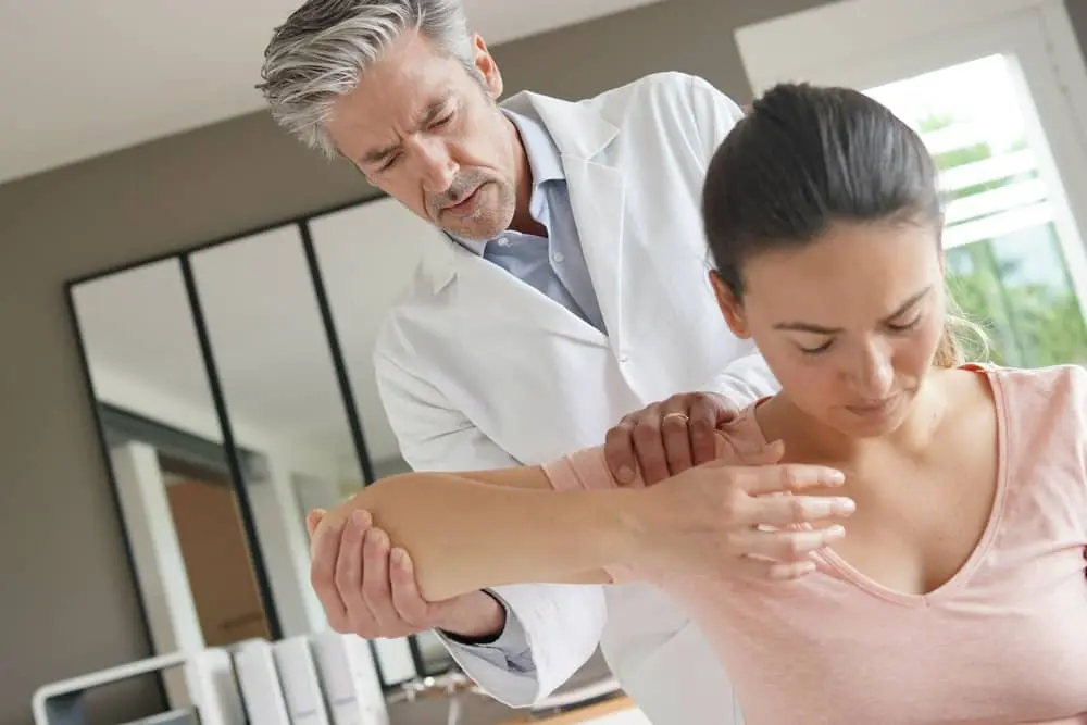
A dislocated shoulder happens when the humerus gets dislodged from the glenoid, causing muscles and tendons to pull out of place and tear. Bone spurs, bony growths in the joint, can grind against tendons, creating friction and possibly a rupture.
Prominent symptoms are pain, decreased range of motion, and instability, which can feel like the shoulder may shift out of place.
The labrum is a fibrous tissue rim surrounding the glenoid (shoulder socket).
A labral tear is a tear found in the labrum. The humerus head is wider than the socket, and the labrum deepens the socket, improving fit and stabilizing the joint. Several ligaments connect this portion of the shoulder. Injuries here might occur due to a single incident or from repetitive use of the shoulder.
A SLAP (known as superior labrum anterior and posterior) tear occurs when a labral tear affects the top part of the socket, where the biceps tendon joins to the shoulder.
Labral tear symptoms include slight clicking or popping noises when you move your arm, catching a sensation in the joint like it can't move any farther. This happens when a piece of labrum gets stuck between the joint bones.
Many people are astonished at how long it takes to repair a soft tissue injury (muscle, tendon, or ligament) and question why they aren't totally recovered and back to normal within two or three weeks.
Unfortunately, this is normal because your body takes much longer to complete tissue recovery.
There are four main stages that your body goes through, although in reality, these aren't distinct, and they all overlap:
Just like a cut to your skin causes external bleeding, a bruise is a sign of bleeding from your internal soft tissues. Muscles have a very good blood supply and hence bleed more and for longer, resulting in a huge bruise. Ligaments have a limited blood supply. Therefore, they bleed less.
Resting during this phase is important to allow the bleeding to stop (approx. 4-6 hours).
Inflammation begins within the first hour or two after injury and peaks within 1-3 days but lasts at least a few weeks. In this phase, you will experience swelling and heat around the injury.
This is completely normal and part of your body's tissue-mending process. It has to happen, and there is nothing you can (or should) do to stop it.
To repair your injury, your body must produce scar tissue. This process begins within 24-48 hours and can extend for several months, usually ending within 4-6 months. So, if you're wondering why you still have some symptoms a couple of weeks after spraining your ankle or knee, it's because your body is busily laying down scar tissue.
The key to helping your body recover during this phase is to gradually exercise in a pain-free way that doesn't overload the brand-new scar but creates a bit of tension within it to build strength and flexibility.
Even when you are past the stages of pain and inflammation, your injury isn't fully recovered. Ligaments, muscles, and tendons have different jobs in your body, and your new scar must be taught to behave like the structure it was formed to repair. Around 2-3 weeks, your body remodels the new scar to be as near the original tissue as feasible. This process can last up to two years.
Your risk of reinjury is higher during this phase due to loss of strength, flexibility, balance, and reaction time, so it's really important to follow a proper rehab program. Your program should also consider any underlying causative factors to prevent recurrence.

Open and close your hand 10-15 times as stated. Then, 10-15 times, move your wrist up and down. Hold a softball/ball of socks after a few days. Squeeze the ball as hard as you can without hurting yourself.
Hold for 5 seconds before repeating ten times.
Bend and straighten your elbow for a mild to moderate stretch. If necessary, use your other arm to assist. Do not force yourself into discomfort.
Raise your elbow to your side. Make a 90-degree bend in it. Slowly spin your palm up and down until a mild to moderate strain is felt. If necessary, use your other arm to assist. Do not force yourself into discomfort.
If symptoms do not worsen, repeat 10-15 times more.
Squeeze the shoulder blades together and bring your shoulders back. You can do this with or without your sling. Hold the position for 20-30 seconds and repeat five times if symptoms do not worsen.
Soft tissue shoulder injuries shouldn’t keep you from functioning normally for too long. Learn more about how experts can help you recuperate and recover from soft tissue shoulder injuries!
Yes. Knee replacement surgery is a major operation, also called arthroplasty, because it uses anesthesia, involves cutting bone, and places permanent implants. Arthroplasty also needs structured rehab for the best long-term result.

Full recovery: Often 3–12 months, depending on health and rehab.
Arthroplasty is a surgery that replaces damaged joint surfaces with artificial parts (implants). In the knee, arthroplasty is used when cartilage loss and joint wear cause pain, swelling, or stiffness that does not improve with non-surgical care.
You may be a candidate for arthroplasty if knee pain limits walking, climbing stairs, sleeping, or basic household tasks. Most people consider arthroplasty after trying options like medication, injections, physical therapy, and activity changes.
If you want an arthroplasty procedure explained in simple terms, think of it as a careful resurfacing of the knee. The surgeon removes the worn areas and restores smoother joint motion using implants.
During knee replacement surgery, the surgeon removes damaged cartilage and a small amount of bone from the joint surfaces. Those surfaces are then shaped to fit the implant.
Here’s what typically happens in arthroplasty:
This is a form of joint replacement surgery, but the steps and rehab plan are knee-specific. The goal is less pain, better movement, and improved stability.

Many patients ask, is knee surgery a major surgery? For knee replacement, the medical answer is yes.
A procedure is often considered major when it involves anesthesia, bone work (cutting and shaping bone surfaces), implants placed inside the body, meaningful blood loss risk, and a recovery period that includes weeks of rehab and physical therapy.
Because knee replacement surgery meets these criteria, it is classified as major. Even though outcomes are often excellent, arthroplasty is still a significant procedure that needs planning and proper aftercare.
Another reason people ask is knee surgery a major surgery is the fear of pain during and after the operation.
For knee replacement surgery, anesthesia is usually spinal anesthesia (numbs the lower body) or general anesthesia (you are asleep). Your team may also use nerve blocks and a structured pain plan to reduce discomfort.
Pain after arthroplasty is expected, but it is also manageable for most patients. The key is early motion, safe walking, and following the recovery plan.
A common question is: how long for knee replacement surgery from start to finish?
Most knee replacement surgery cases take about 1 to 2 hours in the operating room. Complex cases may take longer.
It also helps to know the full day timeline. Even if how long for knee replacement surgery is about two hours, you will spend more time preparing, waking up, and being monitored afterward.
If you are asking how long for knee replacement surgery because you feel nervous, that is normal. Your care team can walk you through the timing step-by-step.

Many people go home the same day after arthroplasty, especially with good support at home. Others stay one night, or sometimes two, depending on pain control, mobility, and medical needs.
Right after knee replacement surgery, most patients will stand and take a few steps with help, start gentle knee movement, learn safe ways to sit and stand, and review home exercises.
This phase is still part of major recovery, which is why joint replacement surgery requires planning. Simple things like food prep, getting a walker, and arranging help at home can make the first week easier.
Recovery after arthroplasty happens in phases. This is where many patients return to the question, is knee surgery a major surgery, because rehab is a real commitment.
Most timelines look like this:
Week 1–2: Swelling and soreness are common. Walking is done with a walker or cane. Home exercises start right away.
Week 3–6: Walking distance and strength improve. Many return to basic daily tasks. Physical therapy stays consistent.
Week 6–12: Knee bend and endurance improve. Stairs feel easier. Many return to driving if cleared.
3–12 months: Strength and confidence continue to build. Movement feels more natural. Final results take time.
Your recovery depends on your health, activity level, and how closely you follow rehab. Arthroplasty results tend to improve steadily with consistent effort.
All major procedures have risks, including joint replacement surgery. Most are uncommon, but it’s important to understand them.
Risks may include infection, blood clots, stiffness, ongoing pain, implant loosening over time, and rare injury to nerves or blood vessels.
Your surgeon will assess your risk factors and explain steps taken to prevent complications. This is one reason arthroplasty planning matters even before surgery day.
Arthroplasty causes post-surgical pain, especially in the first week. Pain usually improves with time, movement, and a structured medication plan. Physical therapy can feel hard early on, but it supports long-term function.
Yes. People often ask is knee surgery a major surgery at age 60, 70, or beyond. The procedure is still major, but many older adults recover well with good preparation, safe mobility, and medical clearance.
For most patients, the main concerns are infection and blood clots. Your team helps reduce these risks with antibiotics, early walking, and prevention plans. Joint replacement surgery is common, but safety still matters.
Most people feel better by 6 to 12 weeks, but full recovery can take months. Even after knee replacement surgery, swelling can linger while strength and endurance rebuild.
Many people return to simple tasks within a few weeks. More demanding activities take longer. After arthroplasty, it’s normal to progress in steps, not all at once.
Knee replacement surgery is major because it involves anesthesia, bone work, implants, and weeks of rehab. Still, for the right patient, arthroplasty can reduce pain and improve function when other treatments stop working.
Have you ever wondered how to effectively treat chronic knee pain or why so many people complain about it today? Chronic knee pain isn't just a fleeting discomfort; it's a persistent aching that lingers, affecting many across age groups. More than just a physical ailment, it's a hurdle that can limit our daily activities. However, hope is still there. At Sunnyvale Orthopedics, we're not just acquainted with this nagging pain; we specialize in understanding its root causes and addressing them head-on. Let's dive deeper, shall we?
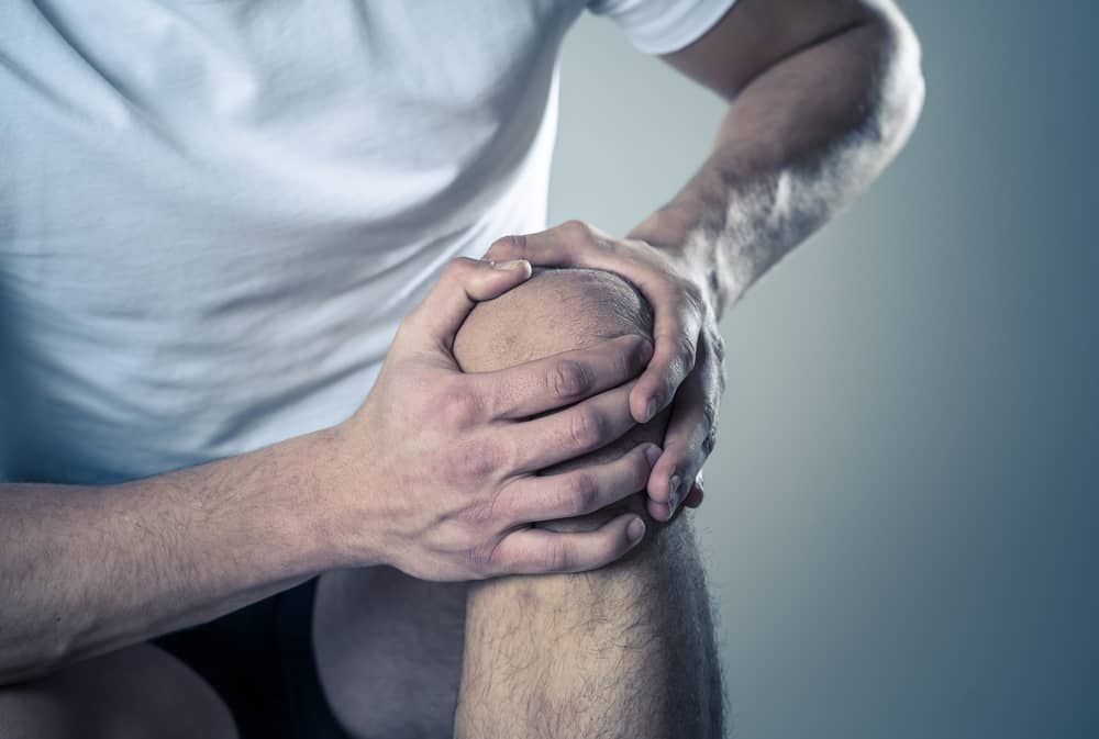
Chronic knee pain is an enduring knee discomfort lasting more than three months. It can stem from various issues such as osteoarthritis, injuries, inflammation, or even wear and tear from age.
While acute knee pain might spring from a sudden injury and tends to heal over time, chronic pain is a constant companion, often making simple tasks like climbing stairs feel like conquering mountains.
Apart from persistent pain, symptoms might include swelling, stiffness, or a popping sound. Sometimes, it's like that uninvited guest who overstays their welcome, affecting your mood and energy.
Imagine wanting to play with your kids or go for that morning jog, but your knees say, "Not today!" Chronic pain can keep you on the sidelines, missing out on cherished moments.
Understanding the intricacies of chronic knee pain is the first step towards finding relief. With a clearer picture of what's happening, you can better navigate your treatment options—especially with the proper guidance.
Before diving into aggressive treatments, it's often best to start with the basics. Conservative treatment methods offer a gentle approach, focusing on non-invasive techniques that can provide significant relief. Let's explore the foundational steps many take to treat chronic knee pain.
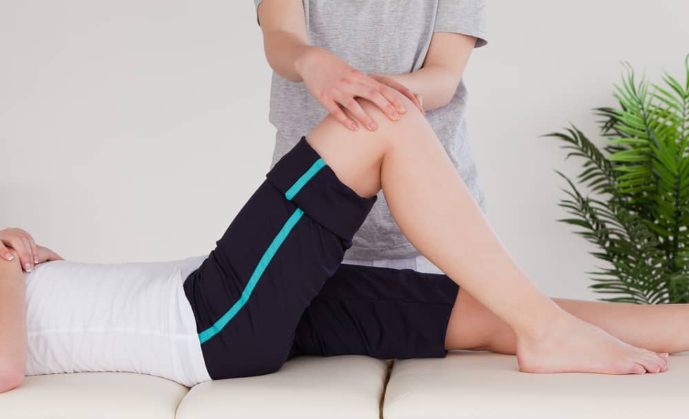
Physical therapy is a cornerstone in knee pain treatment! Focusing on strength and flexibility exercises can work wonders. Let's break it down:
Pain relief can be a game-changer. While we should always be wary and consult professionals:
Think of braces and orthotics as your knee's personal bodyguards. They're here to:
When initial efforts don't bring the desired relief, it's time to consider the next level of intervention. Advanced treatment options delve deeper, employing cutting-edge techniques and technologies to target and treat chronic knee pain aggressively. Here, we'll unravel these sophisticated methods that promise to restore mobility and quality of life.
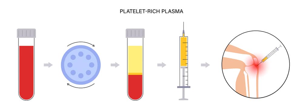
Needles might seem scary, but the relief they bring can be significant. Here's the scoop:
Hyaluronic Acid: It's like lubricating your car but for your joints. This gel-like fluid acts as a cushion and lubricant.
Corticosteroids: These are the big guns for inflammation and pain.
PRP (Platelet-Rich Plasma): Using your body's platelets to promote healing? Genius!
Surgery? But wait, arthroscopy is not your typical surgery. Through tiny incisions, a camera (arthroscope) is inserted to view the inside of the knee. Surgeons can diagnose and treat issues all in one go. Think of it as a knee "tune-up."
Sometimes, despite our best efforts, the natural knee just doesn't want to cooperate. That's where a joint replacement can step in:
By understanding these treatments, you can better navigate your journey to treat chronic knee pain and find the relief you deserve. And always remember, Sunnyvale Orthopedics is here to guide you every step of the way.
Seeking non-traditional methods to treat chronic knee pain? Dive into these natural and alternative therapies that offer a holistic approach to relief, merging ancient wisdom with contemporary practices.
Have you ever had someone tell you about the "magic" of acupuncture? Believe it or not, it's not just magic. Acupuncture and acupressure have roots in ancient Chinese medicine. By targeting specific pressure points, these treatments can help alleviate chronic knee pain. Imagine feeling relief just by the strategic placement of tiny needles or hands-on pressure.
Mother Nature sure has a lot up her sleeve! From turmeric to ginger, several herbs are said to have anti-inflammatory properties. So, the next time you consider treating chronic knee pain, why not give nature a shot? However, a word of caution: always consult a healthcare professional before diving into the herbal world.
"You are what you eat," they say. And it's somewhat true! An anti-inflammatory diet can play a pivotal role in knee pain management. Oh, and shedding those extra pounds? It could be a game-changer. Less weight means less strain on those knees, after all.
Why wait for the pain to become unbearable? Learn the secrets to treat chronic knee pain proactively with these preventive measures and maintenance tips, ensuring your knees stay happy and healthy for the long run.
No, we're not asking you to run a marathon (unless you want to!). But incorporating a routine of low-impact exercises? It's like giving your knees the care they deserve. Regular exercise toughens the muscles around the knee, making it less prone to injury. Think of it as a protective shield.
Have you ever thought about the connection between your shoes and your knees? Well, think about it now. Wearing supportive shoes can make a world of difference. It's like building a house; a strong foundation (or, in this case, shoes) is crucial.
Those niggling signs your knees throw at you? They're like little alarms saying, "Hey! Look here!" Don't ignore them. Recognizing the early symptoms of knee issues and seeking prompt care can make all the difference. It's better to treat chronic knee pain early than to play catch-up later. Remember, it's never too early to care.
Honestly, it's not just about the physical pain. Chronic knee discomfort can drastically limit our life experiences, from simple strolls in the park to dancing at a family wedding. Recognizing and choosing to treat chronic knee pain isn't merely a medical decision; it's a commitment to regaining a life brimming with activity and joy.
So, why wait? Trust the specialists who've made countless folks put their best foot forward. Sunnyvale Orthopedics is here, offering a ray of hope and expert advice.
Have you heard the saying, "Action speaks louder than words"? Well, here's your chance to prove it.
Schedule a consultation at Sunnyvale Orthopedics. Dive deep into the comprehensive strategies we employ to treat chronic knee pain.
With over a quarter-century of experience, Dr. John M. Hibbitts isn't just any orthopedic surgeon. He's your next best ally in your journey towards a pain-free life. Whether it's a worn-out cartilage or a tricky shoulder issue, he and his adept team are your answer. Sunnyvale Sports Medicine and Orthopedic Center: where expertise meets empathy, and challenges transform into success stories.
Act now because of a life without knee pain? It's worth every step.
Frozen shoulder, also known as adhesive capsulitis, is a condition that can significantly impact shoulder mobility without undergoing frozen shoulder treatment. If you are one of the 5-20% of the population suffering from this condition and looking for frozen shoulder treatments, it is important to see the big picture in your journey toward regained shoulder mobility.
In this comprehensive guide, we'll delve into the essential aspects you need to remember before and after your frozen shoulder treatment.
If you're dealing with the discomfort and reduced mobility associated with frozen shoulder, you've likely explored various frozen shoulder treatment options. One of these options includes a highly effective and minimally invasive procedure: Shoulder Arthroscopy.
Shoulder arthroscopy provides a precise and targeted approach to treating frozen shoulder, offering pain relief and restoring its mobility. So, if you want to learn more about this minimally-invasive surgery, this guide will dive deep into shoulder arthroscopy: its benefits, what to keep in mind in preparing for your surgery, and recovering from this effective frozen shoulder treatment.
If you are experiencing persisting indications of shoulder injury, surgery is an option. However, it is generally advised to wait about a year of trying non-invasive frozen shoulder treatments before resorting to surgery since incisions are almost always avoided when possible.
Fortunately, surgery as a frozen shoulder treatment is typically performed as an outpatient procedure—meaning, a patient can go home within 24 hours after the treatment. Thanks to its minimal invasiveness, this frozen shoulder treatment is generally recommended for long-term patients due to its excellent results.
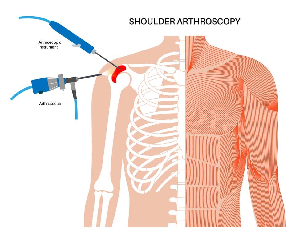
So, what happens during shoulder arthroscopy? During this procedure, the surgeon makes small incisions in your shoulder to remove scar tissue and adjust ligaments, restricting movement. The primary goal of this frozen shoulder treatment is to release the tight and inflamed capsule around the shoulder joint, allowing for better shoulder mobility.
This is done using a small tool called an arthroscope, equipped with a camera and light source, inserted through tiny shoulder incisions. The camera sends real-time images to a monitor, giving the surgeon a precise view of the shoulder joint.
Since this frozen shoulder treatment is minimally invasive, it often leads to a quicker recovery and potentially higher success rates by minimizing damage to the surrounding connective tissue. Smaller surgical instruments are used, and the ability to see the joint on a video monitor allows for prompt and accurate diagnosis and repair of any damaged joint tissue. This procedure ultimately aims to unlock your frozen shoulder and regain its full range of mobility.
When it comes to ensuring your upcoming frozen shoulder treatment goes smoothly, it goes without saying that following your physicians’ advice is paramount. But aside from your shoulder pain doctor’s advice, here are some general reminders before your frozen shoulder treatment:
Although shoulder arthroscopy is minimally invasive, your shoulder joint still needs weeks to months to heal completely. After your frozen shoulder treatment, it is normal to experience some pain and swelling for at least several weeks.
This is where a rehabilitation plan comes in. It includes gentle exercise and physical therapy, which ultimately aim to increase your shoulder movement and strength over time. Based on your lifestyle and frozen shoulder treatment, your healthcare provider will give you a rehabilitation plan specific to you.

Here is what to expect after your frozen shoulder treatment:
With all these precautions in mind, it is easy to be overwhelmed with your shoulder’s recovery process. However, remember that the weeks of recovery can last you long-term relief from stiff and immobile shoulder, letting you experience pain-free everyday activities.
While prevention is often deemed the best cure, the importance of follow-up care is just as crucial to keeping your shoulders healthy. After your frozen shoulder treatment, your surgeon will schedule follow-up appointments to monitor your progress. These check-ups are where your surgeon can monitor your healing progress, as well as assess if there are necessary adjustments to your treatment and rehabilitation plan.
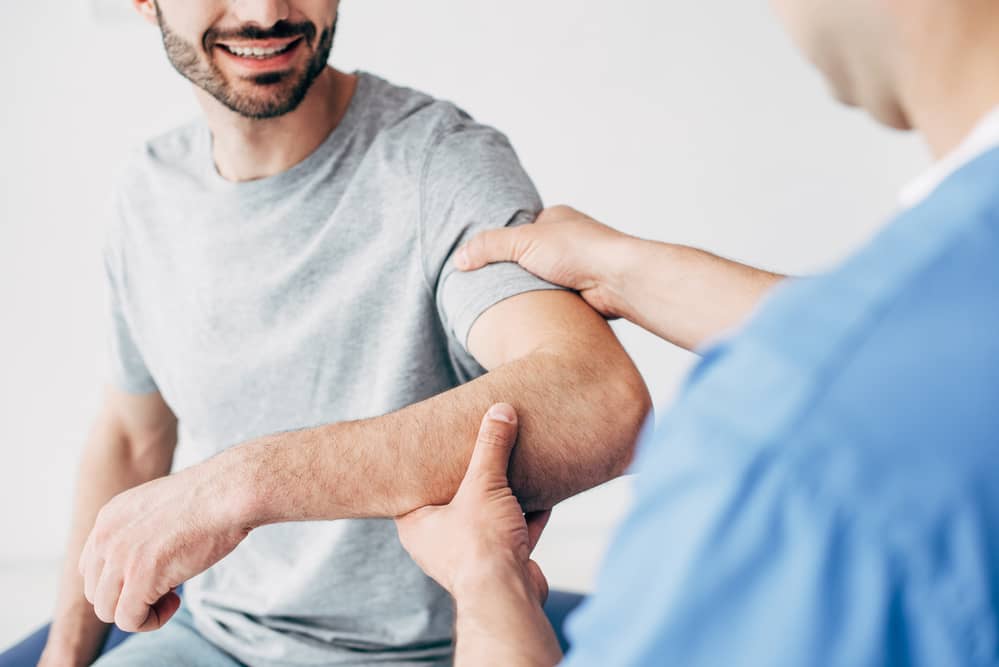
Bear in mind that the road to recovery after shoulder arthroscopy is gradual. Avoid straining your shoulder, and be patient during the healing process. The weeks of recovery may seem like a long time now, but you will see how taking your time to slow down is worth all the pain-free days in the future.
With diligent medical care, supervision, and proper maintenance, your frozen shoulder treatment has the potential to restore your shoulder to its full functionality. Remember to always consult with and adhere to your doctor's guidance for a smooth and successful recovery journey.
The shoulder is like a ball and socket connection. When the arm bone's ball part (humerus) is pushed out of the shoulder's socket (glenoid), it's called shoulder instability or dislocation. This often happens from injury, but it can also be caused by doing the same movements too much in sports like baseball or swimming. When the shoulder gets partly or fully dislocated, it can hurt and limit how well it works. This leads us to the question: what causes shoulder instability, and can surgery fix shoulder instability?
While the shoulder has an impressive range of motion, it can still lose its stability. The following are types of shoulder instability:
With significant trauma to a previously normal joint, the humeral head can be forcefully subluxated or dislocated. A shoulder subluxation happens when the humerus partially slides in and out of place quickly. Shoulder dislocations occur when the humerus comes out of the glenoid. It may fall back into place after time or may need to be put back into place with medical assistance.
During shoulder subluxation and dislocation, the capsule, ligaments, or labrum can be stretched, torn, or detached from the bone. Once the humeral head is back in place, these structures can heal in a loose or stretched position that may increase the risk of future episodes of subluxation or dislocation. With each additional episode, more tissue damage can occur, increasing the tendency toward future instability.
Shoulder instability can occur when the labrum is torn or peeled off the glenoid. This can occur after a shoulder dislocation, trauma, or repetitive motion (like throwing a baseball).
Some individuals are born with somewhat loose shoulder ligaments (they have a loose or spacious capsule). For these people, instability can occur without trauma or following relatively minor injury. Some patients may also have a genetic condition that causes looseness in the joints and predisposes them to develop shoulder instability or weakness.
People with the instability of the shoulder joint can often feel the ball of the shoulder come out of its socket or "give way." It's commonly associated with pain. Typically, the episodes of giving way happen with specific activities or positions of the arm, like throwing a ball or reaching behind the body.
More symptoms can include a decreased range of arm/shoulder motion, swelling, and bruising.
An orthopedic doctor should do a complete history and physical examination. The examination includes palpation for checking points of tenderness and determining range of motion and strength. Specific tests can also assess the laxity of the shoulder joint or the degree of shoulder looseness during the examination. X-rays are usually done to determine the possible causes of the instability and rule out other causes of shoulder pain, like a fracture.
Additional tests, like a magnetic resonance imaging (MRI) scan or a dye test (arthrogram) with or without a computed tomography (CT) scan, are occasionally done further to evaluate the bones and tissues of the shoulder joint. However, these scans are not required in all patients with instability.
After a shoulder has dislocated or subluxed, it's important to rest it and avoid aggravating activities for a few days. If the shoulder pain is significant, like following a traumatic dislocation, a sling is usually used to provide temporary immobilization — shoulder bracing may also be an option for some patients. Range of motion exercises are started once the pain and swelling have subsided. Strengthening exercises can begin as motion improves. The exercise program is typically done with a trained physical therapist.
Cold packs or ice bags on your shoulder before and after working out can help lessen pain and swelling. Medications called NSAIDs, which include aspirin, ibuprofen (found in brands like Motrin, Advil, and Nuprin), or similar drugs like Aleve, can also help decrease pain and swelling. You should check with your physician with any questions, as several different kinds of drugs are available, and they may have different side effects.
Therapy helps get your shoulder moving again and makes the muscles around it stronger. These muscles, especially the ones in the rotator cuff, must be strong to keep your shoulder from dislocating or partially coming out of place. When your shoulder is working well, you can start doing your usual activities bit by bit.
Yes, it can! Nonetheless, the procedure(s) that orthopedic surgeons will advise to treat shoulder instability depends on factors like:
When a shoulder moves out of place towards the front of the body, it can tear the cartilage ring (called the labrum) surrounding the socket.
This is known as a Bankart lesion. A Bankart repair aims to fix the tear in the labrum and attach it back to the socket. This repair can be done through traditional open surgery or a less invasive arthroscopic method.
Especially after multiple dislocations, the ligaments holding the shoulder joint in place can become stretched and lax. A capsular shift makes the ligaments tighter by making flaps in the tissue, moving them, and sewing them again to remove any looseness.
The Latarjet procedure (otherwise known as Bristow or Bristow-Latarjet procedure) can be utilized when a bone on the edge of the socket gets damaged during a dislocation. In this treatment, bone is moved from the coracoid process (a part of the shoulder blade) along with the attached conjoint tendon to the front edge of the socket. The Latarjet procedure can be performed through open surgery or arthroscopy.
The remplissage procedure can be performed along with the Latarjet procedure. It might be suggested if the ball of the shoulder, also called the humeral head, is seriously compromised due to dislocation, which results in a problem called Hill-Sachs lesion. This procedure includes repairing the head's surface and filling the gap by attaching it to a section of the rotator cuff.

The ball in the shoulder might need to be replaced (hemiarthroplasty), or both the ball and socket might need to be replaced (total shoulder arthroplasty) to fix shoulder problems. Sometimes, if the ball side has big bone defects that can't be fixed with a bone graft, hemiarthroplasty is needed to rebuild it.
Older patients might have shoulder issues when their shoulder becomes unstable. Sometimes, big tears in the shoulder's rotator cuff can't be fixed. For these cases, a reverse shoulder replacement could be the best option. This involves putting a ball in the socket and attaching a plastic socket to the ball using a stem in the arm bone.
Even after physical therapy to regain full shoulder movement and strength, the shoulder might feel unsafe or unstable. So, there are two main choices for treatment: 1) changing how you do things and 2) having an operation. Changing how you do things is mostly for people who feel their shoulder isn't steady, only when they do specific activities like playing basketball or sports with overhead racquets.
In these patients, avoiding the activity can eliminate their episodes of subluxation or dislocation.
Some patients might need surgery if they don't want to quit activities or sports that set off their issues. Surgery is also an option for those whose routine tasks like dressing or sleeping become tough due to instability.
The surgery includes an examination of the shoulder under anesthesia to fully assess the extent and direction of the instability while the muscles surrounding the shoulder are completely relaxed. An arthroscope is frequently used to inspect the inside of the shoulder joint to evaluate the joint and its cartilage. The arthroscope allows direct assessment of the condition of the labrum and rotator cuff tendons. In a limited number of select patients with relatively mild looseness or laxity, it may be possible to stabilize the shoulder by arthroscopic techniques.
To correct severe instability, open surgery is often necessary. An incision is made over the shoulder, and the muscles are moved to gain access to the joint capsule, ligaments, and labrum.
Depending on the tissue injury identified at surgery, these structures are either repaired, reattached, or tightened. The repair can be done with simple sutures attached to metal or secured to plastic or absorbable tacks or anchors. These anchors are inserted into the bone and hold the sutures used to reattach or tighten the ligaments. These anchors stay in the bone permanently.
The course of recovery following surgery depends upon the type of procedure the surgeon performs. Usually, the hand, wrist, and elbow range of motion begins the day after surgery. Most patients can write and use their arms to eat within three to seven days after surgery. A supervised physical therapy program is initiated one to four weeks after the operation. A full range of motion usually returns after six to eight weeks. Strength usually returns in three months.
Driving sometimes takes several weeks. Return to work or sporting activities depends on the specific nature and demands of that activity but can take up to one year or more for heavy laborers or high-level athletes. With surgery, the chance of recurrence of the instability is low (3 percent to 5 percent), and most patients can go back to their previous activities.
Many of us only notice our shoulder joints' importance when they get hurt. Then, we feel pain and other problems that disrupt what we usually do. To prevent long-lasting pain and keep our shoulder joints healthy, it's best to check shoulder pain early when simple treatments work well. If your shoulder hurts, don't wait. Contact us at Sunnyvale Sports Medicine and Orthopedic Center!
Proper diagnosis is needed to provide accurate, broken ankle treatment to individuals. Ankle injuries are the most prevalent type of bone and joint injury. Often, the degree of discomfort, incapacity to walk, or fear that a bone has been shattered prompts you to seek emergency care. However, it is essential to note that not all ankle injuries require immediate medical attention. Rest, ice, compression, and elevation (RICE) can help alleviate pain and swelling in some situations. Consulting a healthcare professional for a proper diagnosis and treatment plan is always advisable.
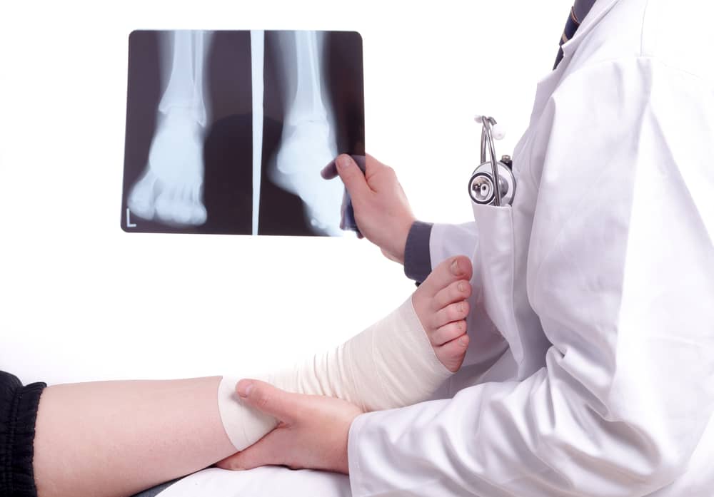
Your main concern is the same as the doctor's: Is there a broken bone? Without ankle X-rays, distinguishing between a fracture (broken bone) and a sprain, dislocation, or tendon injury is often impossible. Ankle X-rays are critical for assessing the degree of an injury and making an accurate diagnosis and broken ankle treatment. Depending on the severity of the injury, a healthcare practitioner can prescribe the best course of treatment, which could include immobilization, physical therapy, or surgery.
An ankle joint is injured when it is stressed beyond the strength of its parts. Sprained ankle occurs when only the ligaments give way and tear. An ankle fracture occurs when a bone loses way and breaks.
Fractures can develop in conjunction with ligament rips. When too much force is applied to the ankle, it breaks. The most common causes are:
Falls and slips. Losing your equilibrium might cause you to trip and fall, putting too much weight on your ankle. This can happen if you walk on an uneven surface, wear ill-fitting shoes, or wander around in the dark. High-impact exercises or rapid movements can cause excessive stress on the ankle joint, resulting in a break. To limit the incidence of ankle fractures, it is critical to take preventative steps and wear suitable protective gear when participating in strenuous activities.
Significant impact. A broken ankle treatment might be needed if a situation arise from the force of a jump or fall. Even if you jump from a short height, this can happen. The extremity of a broken ankle depends on the force of the impact. A slight fracture may occur in certain circumstances, while the ankle may be destroyed in others. As a result, it is critical to be vigilant and take the required steps to avoid any potential mishaps or injuries.
Missteps. If you place your foot incorrectly, you risk breaking your ankle. It may twist or roll to the side as you put weight on your ankle. This can cause your ankle bones to fracture or dislocate. In addition, high-impact activities such as sports or jumping from great heights might increase the likelihood of a mishap and subsequent ankle injury. To reduce the danger of a broken ankle, it is critical to exhibit caution and employ good technique when participating in these sports.
Sports. High-impact sports involve intense movements that stress the joints, including the ankle. Examples of high-impact sports include soccer, football, and basketball. These sports frequently entail abrupt changes in direction, rapid accelerations, and jumping, which can place undue strain on the ankle joint. To lessen the risk of an ankle injury, it is critical to wear appropriate footwear with excellent ankle support and to warm up properly before indulging in these activities.
Car accidents. A car accident's sudden, hard impact might result in shattered ankles. These injuries frequently necessitate surgical treatment. To reduce the risk of car accidents, it is vital to practice safe driving behaviors, follow traffic rules, wear appropriate footwear, and warm up correctly. However, if a car accident occurs and an ankle injury is inflicted, it is critical to seek prompt medical assistance to ensure appropriate diagnosis and treatment.
Overuse. A stress fracture, a microscopic split in the bone, can be caused by repetitive strain or damage to the ankle. This is a common injury among runners and other athletes. Listening to your body and allowing enough rest and recovery time is critical to avoid overuse problems. Wearing supportive footwear and employing orthotics can also help lower the risk of stress fractures in the ankle.
Trauma. Crush damage and fracture can result from an intense impact on the ankle. A significant object falling on the foot or heavy machinery running over the ankle could cause this. Ankle injuries can occur due to rapid twists or rolls of the ankle and trauma. These injuries can happen when participating in sports such as running, leaping, or even walking on uneven surfaces.
Direct trauma, such as a fall or impact during sports activities, is another prevalent cause of ankle fractures. Fractures can also occur due to underlying disorders such as osteoporosis, which weakens bones and makes them more prone to breaking.
Your doctor will inspect your ankle to look for sensitive spots. The particular location of your pain might aid in determining its origin.
Your doctor may move your foot into various positions to assess your range of motion. Your doctor may ask you to walk for a short distance so that he or she can assess your gait.
Based on the results of these physical examinations, your doctor may also order additional tests such as X-rays or MRI scans to further evaluate the condition of your ankle. These diagnostic tests can provide more detailed information about potential fractures, ligament tears, or other underlying conditions that may be causing your pain and affecting your mobility.
Your doctor might suggest one or more imaging tests listed below if your symptoms and signs point to a break or fracture.
X-rays. X-rays can show the majority of ankle fractures. The technician may need to take X-rays from several angles to ensure that the bone pictures do not overlap too much. Stress fractures frequently do not show up on X-rays until the break begins to mend.
Scan of the bones. A bone scan can assist your doctor in diagnosing fractures that are not visible on X-rays. The doctor will inject a small amount of radioactive material into a vein. The radioactive material is drawn to your bones, particularly the damaged areas of your bones. Damaged areas, such as stress fractures, appear as bright spots in the final image. A bone scan helps detect and diagnose stress fractures that X-rays may miss. In contrast to X-rays, which simply show the structure of the bones, a bone scan can disclose the extent of damage and pinpoint specific locations that require care. The scan shows any abnormalities in your bones by infusing a small amount of radioactive material into your bloodstream, allowing your doctor to accurately identify and treat your illness.
Computed Tomography Scans. CT scans X-rays from various angles and combines them to create cross-sectional images of your body's internal structures. CT scans can give more information about the injured bone and the surrounding soft tissues. A CT scan may assist your doctor in determining the best course of treatment for your broken ankle.
Magnetic Resonance Imaging. Magnetic resonance imaging (MRI) creates incredibly detailed images of the ligaments holding your ankle together using radio waves and a strong magnetic field. This imaging aids in identifying fractures and ligaments that are not visible on X-rays.
Ankle fracture treatment is determined by factors such as the number of shattered ankle bones. Although not all broken ankles necessitate surgery, all broken ankles need some level of care to heal.
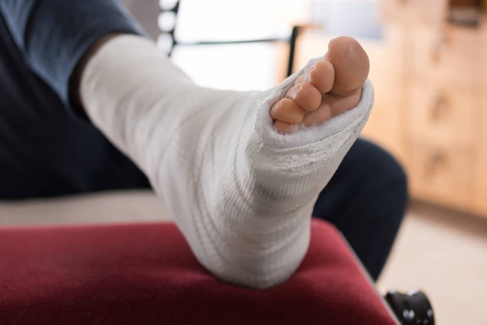
If you have a stress fracture, you probably won't need surgery. A brace or cast may be required to support your ankle while it heals. More severe ankle fractures necessitate reduction or surgery.
Reduction is how healthcare personnel adjust your ankle to align the ends of your shattered bones.