(214) 420-7611


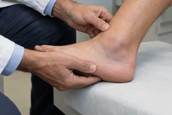
Did you roll your ankle and are wondering how bad the injury is? The sprained ankle vs broken ankle question isn’t always easy to answer. A sprain affects the ligaments, while a fracture means a bone has cracked or broken. Both can leave your ankle swollen, bruised, and painful.
The American Academy of Orthopaedic Surgeons (AAOS) notes that a severe sprain can cause pain, bruising, and swelling similar to a fracture, which is why the two are easy to confuse.
Both injuries are common. US emergency departments treated an estimated 7.4 million ankle sprains from 2010 to 2024, and the AAOS lists ankle fractures as the fourth most common type of fracture in adults.
You might still be able to walk with a fracture. That’s why it’s worth getting checked if you have tenderness over the ankle bones or can’t take four steps.
An ankle sprain happens when the ligaments supporting your joint stretch too far or tear. A fracture involves one or more bones around the ankle, usually the tibia, fibula, or talus.
Around 90% of ankle sprains are inversion injuries, where the foot rolls inward and damages the ligaments on the outside of the ankle. Doctors grade sprains by how much the ligament is damaged:
• Grade 1: Slight stretching and microscopic tearing of the ligament fibers, with mild swelling and usually no pain when standing.
• Grade 2: A partial tear, with moderate swelling, mild pain when standing, and slight looseness in the joint.
• Grade 3: A complete tear, with significant swelling, severe pain when standing, and a lot of instability.
Fractures are described by which part of the bone breaks. A break at the lower end of the fibula, the bony bump on the outer ankle, is a lateral malleolus fracture. When the bones break on both the inner and outer sides, it’s a bimalleolar fracture. A trimalleolar fracture also involves the back of the tibia. Doctors also note whether the bone pieces have shifted out of place (displaced) or stayed in line (nondisplaced).
The two injuries can also overlap. Some fractures happen together with torn ligaments. A high ankle sprain damages the ligaments that hold the lower tibia and fibula together. The AAOS says these sprains can usually be treated without surgery, but they may take longer to heal than a typical ankle sprain.
Here’s how the two injuries compare.
| Feature | Sprained Ankle | Broken Ankle |
| What’s injured | Ligaments | Bones |
| Common cause | Rolling or twisting | Falls, twists, or impact |
| Pain | Mild to severe | Mild to severe |
| Swelling | Common | Common |
| Walking | May be difficult | May still be possible |
| Ankle shape | Usually normal despite swelling | May look crooked |
Hearing a pop doesn’t mean you’ve broken a bone. Ligaments can make a similar sound when they tear. According to the AAOS, you may hear or feel a pop when the ligaments tear severely.
A broken ankle doesn’t always look as bad as you’d expect. Some fractures cause intense pain right away. Others feel more like a bad sprain. The AAOS describes a wide range, from a simple break in one bone that may not stop you from walking to several breaks that may need one or more surgeries.
Watch for these broken ankle symptoms:
Seek emergency care if your ankle looks deformed, bone is visible, or your foot feels cold or numb. When a broken bone pushes through the skin, it’s called an open fracture. The AAOS treats it as an emergency because dirt and debris can enter the wound and raise the risk of infection.
Yes, you can walk on a broken ankle in some cases. Small, stable fractures may allow you to stand or limp around. The AAOS notes that many isolated fractures at the end of the fibula are stable enough for doctors to allow weight on the ankle during treatment.
That doesn’t mean walking is safe. Putting weight on an injured ankle could make an unstable fracture worse.
If the weight goes on a fracture too early, the bone pieces can move out of place. The bone may then heal in the wrong position (malunion) or not heal at all (nonunion).
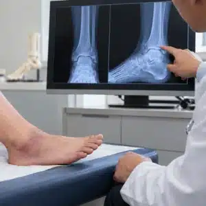
To check stability, doctors may order a stress X-ray, which is taken while the ankle is under pressure or bearing weight. It helps show whether a fracture is stable or more likely to need surgery.
A severe sprain can also make walking impossible. Your ability to stand isn’t enough to tell the two injuries apart.
Under the Ottawa Ankle Rules, which many doctors use to decide on X-rays, limping still counts as bearing weight. That’s why the exam also checks for tenderness over specific bones. Bone tenderness alone can be enough to call for an X-ray.
Sometimes—but being able to walk does not mean it is safe.
Small, stable fractures may still allow standing or limping.
Early weight on an unstable fracture can shift bones and affect healing.
Bone tenderness may warrant an X-ray—even if you can walk.
Walking does not rule out a fracture. Severe sprains can also prevent walking.
Not every twisted ankle needs an X-ray. Doctors often use the Ottawa Ankle Rules to decide whether imaging is necessary.
Emergency physicians at the University of Ottawa developed the rules in 1992 to cut unnecessary imaging, since fewer than 15% of people X-rayed for an ankle injury in emergency departments have a fracture.
A review published in the BMJ found these rules could reduce unnecessary X-rays by roughly 30% to 40%.
The 2003 review pooled 27 studies involving 15,581 adults and children. It found the rules have a sensitivity of almost 100%, which means they rarely miss a fracture. In most groups studied, fewer than 2% of patients who didn’t meet the X-ray criteria actually had an ankle or midfoot fracture.
An ankle X-ray is generally recommended when pain near the ankle bones comes with either:
For pain in the middle of the foot, doctors check two other areas: the navicular bone and the base of the fifth metatarsal. Tenderness there, or the same four-step problem, may call for a foot X-ray.
The navicular sits on the inner side of the midfoot, while the base of the fifth metatarsal is the bump on the outer edge of the foot. Dr. Ian Stiell, who led the development of the rules, notes that patients rarely need both an ankle and a foot X-ray. Usually, one or the other is enough, if any.
These rules aren’t suitable for every patient. Young children and people who cannot give a reliable response to an exam may need a different approach. The rules’ creators advise caution in patients under 18. They also recommend clinical judgment when an exam may be unreliable, such as with intoxication, other painful injuries, reduced feeling in the legs, or swelling too severe to feel the bones.
An X-ray can reveal a fracture, but it won’t show most ligament damage clearly. Your doctor may recommend other scans if symptoms don’t improve.
According to the AAOS, an MRI can show ligaments, cartilage, and tendons. It may be ordered for a suspected high ankle sprain or when symptoms last beyond 6 to 8 weeks despite treatment. A CT scan gives a more detailed view of the bones and can help with surgical planning.
The first thing to do is stop whatever caused the injury. Don’t try to walk it off when you’re in significant pain.
While arranging an exam, you can:
Once a fracture has been ruled out, gentle movement and walking as tolerated often help sprains recover. Your doctor can advise when to start.
The AAOS notes that early weight-bearing as tolerated is typically recommended for sprains and that ending immobilization early in rehab helps prevent stiffness. In most cases, sprain pain and swelling last 2 to 3 days, though walking may be difficult during that time.

Treatment depends on what was injured and how badly.
For ankle sprains, a brace and physical therapy are often enough. Exercises help restore movement, strength, and balance. Surgery is uncommon, even for serious sprains.
The AAOS says nearly all isolated low ankle sprains can be treated without surgery, and even a complete tear can heal with proper immobilization and rehab. A grade 2 sprain may call for a walking boot or brace. A grade 3 sprain may need a short leg cast or cast-brace for 10 to 14 days. Rehab can take as little as 2 weeks for minor sprains and up to 6 to 12 weeks for more severe injuries.
Finishing rehab matters. The AAOS identifies incomplete rehabilitation as the most common cause of chronic ankle instability, a condition where the ankle keeps giving way. In one study that followed 82 people after a first-time sprain on the outer ankle, 40% had chronic ankle instability 12 months later. Balance training can also help prevent repeat sprains. In a 2009 BMJ trial of 522 athletes, an 8-week home balance program reduced the risk of another sprain by 35%. Repeat sprains occurred in 22% of athletes who did the program, compared with 33% of those who didn’t.
For broken ankles, stable fractures may heal in a cast or walking boot. Bones that have shifted out of place may need surgery.
Surgeons often repair displaced fractures with screws, a plate, and screws, or a rod. According to the AAOS, most ankle fractures that come with a dislocation need surgery, as do most bimalleolar fractures. People who smoke, have diabetes, or are older face a higher risk of complications after surgery.
The American Academy of Orthopedic Surgeons (AAOS) notes that ankle fractures typically need at least six weeks for early bone healing. Complete bone healing may take 10 to 12 weeks.
Getting back to sports or other demanding activities can take longer. The AAOS says most people return to normal activities within 3 to 4 months. If the right ankle is affected, driving can often resume at 9 to 12 weeks. Some people still limp after several months, stiffness and swelling can linger even after the bone heals, and complete recovery from some ankle fractures can take up to 2 years.
Doctors have more tools to assess injuries and guide recovery, though not every clinic uses them.
AI-assisted X-rays: A 2026 review in Skeletal Radiology looked at how well AI could spot broken bones in foot and ankle X-rays. The results were encouraging, but doctors still need to read the scans themselves. Researchers also want to see how these tools perform in everyday clinics.
A 2026 study from Geneva University Hospitals shows why. Among 701 adults with foot or ankle injuries, musculoskeletal radiologists caught 84% of fractures, compared with 74.3% for one commercial AI tool working on its own. The radiologists also did better with complex midfoot fractures.
Home-based rehabilitation: Recent studies have also looked at remote exercise programs for people with ongoing ankle instability. Guided home exercises may help selected patients improve their balance and movement. They aren’t a replacement for examining a possible fracture.
It’s difficult because both injuries can cause swelling, bruising, and pain. Tenderness over the ankle bones or trouble taking four steps raises concern for a fracture. A medical exam can help determine whether you need an X-ray.
Yes, some people can walk with a small, stable fracture. They may assume it’s only a sprain because the ankle still supports their weight. If you have pain directly over the bone, get it examined promptly.
No. If you can walk and don’t have tenderness over certain bones, you may not need an X-ray. Your doctor will check the injury and use the Ottawa Ankle Rules to help make that decision.
Early bone healing usually takes at least six weeks. The AAOS says complete healing often takes 10 to 12 weeks. Regaining your usual strength and movement may take several more months.
It can. A badly torn ligament may leave your ankle feeling weak or unsteady long after the swelling goes down. Meanwhile, a small fracture that stays in place may heal well with a cast or boot.
That can happen, especially if you've hurt a ligament rather than a bone. Some small fractures are also hard to see on the first X-ray. If the pain isn't getting better, have your ankle checked again.
A twisted ankle might seem minor, but the right diagnosis can prevent bigger problems later. Sunnyvale Sports Medicine & Orthopedic Center helps patients in Sunnyvale and nearby East Dallas communities with ankle injuries. Still unsure what's causing your pain? Book an ankle evaluation and find out what care you need.